Parvalbumin

Last updated
PVALB
Available structures
PDB Ortholog search: PDBe RCSB
Identifiers
Aliases PVALB , D22S749, parvalbumin
External IDs OMIM: 168890 MGI: 97821 HomoloGene: 2137 GeneCards: PVALB
Orthologs
SpeciesHumanMouse
Entrez
Ensembl
UniProt
RefSeq (mRNA)

NM_002854
NM_001315532

NM_013645
NM_001330686

RefSeq (protein)

NP_001302461
NP_002845
NP_001302461.1
NP_002845.1

NP_001317615
NP_038673

Location (UCSC) Chr 22: 36.8 – 36.82 Mb Chr 15: 78.08 – 78.09 Mb
PubMed search [3] [4]
Wikidata
View/Edit Human View/Edit Mouse
Parvalbumin
Identifiers
Symbol?
InterPro IPR008080

Parvalbumin (PV) is a calcium-binding protein with low molecular weight (typically 9-11 kDa). In humans, it is encoded by the PVALB gene. It is a member of the albumin family; it is named for its size (parv-, from Latin parvus which means "small") and its ability to coagulate.

Contents

It has three EF hand motifs and is structurally related to calmodulin and troponin C. Parvalbumin is found in fast-contracting muscles, where its levels are highest, as well as in the brain and some endocrine tissues.

Parvalbumin is a small, stable protein containing EF-hand type calcium binding sites. It is involved in calcium signaling. Typically, this protein is broken into three domains, domains AB, CD and EF, each individually containing a helix-loop-helix motif. [5] The AB domain houses a two amino-acid deletion in the loop region, whereas domains CD and EF contain the N-terminal and C-terminal, respectively. [5]

Calcium binding proteins like parvalbumin play a role in many physiological processes, namely cell-cycle regulation, second messenger production, muscle contraction, organization of microtubules and phototransduction. [6] Therefore, calcium-binding proteins must distinguish calcium in the presence of high concentrations of other metal ions. The mechanism for the calcium selectivity has been extensively studied. [6] [7]

Location and function

Pvalb is expressed in the reticular nucleus of the thalamus in the postnatal day 56 mouse. Allen Brain Atlases Pvalb, mouse, ret thalamus, ISH.jpg
Pvalb is expressed in the reticular nucleus of the thalamus in the postnatal day 56 mouse. Allen Brain Atlases
In the cerebellum of adult mice Pvalb is expressed in Purkinje cells and molecular layer interneurons. Allen Brain Atlases Pvalb, cerebellum, ISH.jpg
In the cerebellum of adult mice Pvalb is expressed in Purkinje cells and molecular layer interneurons. Allen Brain Atlases

In neural tissue

Parvalbumin is present in some GABAergic interneurons in the nervous system, especially the reticular thalamus, [8] and expressed predominantly by chandelier and basket cells in the cortex. In the cerebellum, PV is expressed in Purkinje cells and molecular layer interneurons. [9] In the hippocampus, PV+ interneurons are subdivided into basket, axo-axonic, and bistratified cells, each subtype targeting distinct compartments of pyramidal cells. [10]

PV interneurons' connections are mostly perisomatic (around the cell body of neurons). Most of the PV interneurons are fast-spiking. They are also thought to give rise to gamma waves recorded in EEG.

PV-expressing interneurons represent approximately 25% of GABAergic cells in the primate DLPFC. [11] [12] Other calcium-binding protein markers are calretinin (most abundant subtype in DLPFC, about 50%) and calbindin. Interneurons are also divided into subgroups by the expression of neuropeptides such as somatostatin, neuropeptide Y, cholecystokinin.

In muscular tissue

PV is known to be involved in relaxation of fast-twitch muscle fibers. [13] [14] This function is associated with PV role in calcium sequestration.

During muscle contraction, the action potential stimulate voltage-sensitive proteins in T-tubules membrane. These proteins stimulate the opening of Ca2+ channels in the sarcoplasmic reticulum, leading to release of Ca2+ in the sarcoplasm. The Ca2+ ions bind to troponin, which causes the displacement of tropomyosin, a protein that prevents myosin walking along actin. The displacement of tropomyosin exposes the myosin-binding sites on actin, permitting muscle contraction. [15]

This way, while muscle contraction is driven by Ca2+ release, muscle relaxation is driven by Ca2+ removal from sarcoplasm. Along with Ca2+ pumps, PV contributes to Ca2+ removal from cytoplasm: PV binds to Ca2+ ions in the sarcoplasm, and then shuttles it to the sarcoplasmic reticulum. [16]

Clinical significance

Decreased PV and GAD67 expression was found in PV+ GABAergic interneurons in schizophrenia. [17] [18]

Parvalbumin has been identified as an allergen causing fish allergy (but not shellfish allergy). [19] [20] [21] [22] Bony fishes manifest β-parvalbumin and cartilaginous fishes such as sharks and rays manifest α-parvalbumin; allergenicity to bony fishes has a low cross-reactivity to cartilaginous fishes. [20]

History

The protein was discovered in 1965 as a component of the fast-twitching white muscle of fish. It was described as a low molecular-weight "albumin". [23] It is unknown who coined the term parvalbumin, but the word is already in use by 1967. [24]

Related Research Articles

<span class="mw-page-title-main">Calmodulin</span> Messenger protein

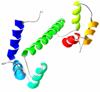
Calmodulin (CaM) (an abbreviation for calcium-modulated protein) is a multifunctional intermediate calcium-binding messenger protein expressed in all eukaryotic cells. It is an intracellular target of the secondary messenger Ca2+, and the binding of Ca2+ is required for the activation of calmodulin. Once bound to Ca2+, calmodulin acts as part of a calcium signal transduction pathway by modifying its interactions with various target proteins such as kinases or phosphatases.

<span class="mw-page-title-main">Interneuron</span> Neurons that are not motor or sensory

Interneurons are neurons that connect to brain regions, i.e. not direct motor neurons or sensory neurons. Interneurons are the central nodes of neural circuits, enabling communication between sensory or motor neurons and the central nervous system (CNS). They play vital roles in reflexes, neuronal oscillations, and neurogenesis in the adult mammalian brain.

Voltage-gated calcium channels (VGCCs), also known as voltage-dependent calcium channels (VDCCs), are a group of voltage-gated ion channels found in the membrane of excitable cells (e.g., muscle, glial cells, neurons, etc.) with a permeability to the calcium ion Ca2+. These channels are slightly permeable to sodium ions, so they are also called Ca2+–Na+ channels, but their permeability to calcium is about 1000-fold greater than to sodium under normal physiological conditions.
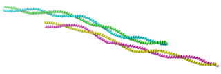
<span class="mw-page-title-main">Calcium signaling</span> Intracellular communication process

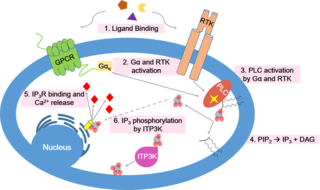
Calcium signaling is the use of calcium ions (Ca2+) to communicate and drive intracellular processes often as a step in signal transduction. Ca2+ is important for cellular signalling, for once it enters the cytosol of the cytoplasm it exerts allosteric regulatory effects on many enzymes and proteins. Ca2+ can act in signal transduction resulting from activation of ion channels or as a second messenger caused by indirect signal transduction pathways such as G protein-coupled receptors.

<span class="mw-page-title-main">Glutamate receptor</span> Cell-surface proteins that bind glutamate and trigger changes which influence the behavior of cells

Glutamate receptors are synaptic and non synaptic receptors located primarily on the membranes of neuronal and glial cells. Glutamate is abundant in the human body, but particularly in the nervous system and especially prominent in the human brain where it is the body's most prominent neurotransmitter, the brain's main excitatory neurotransmitter, and also the precursor for GABA, the brain's main inhibitory neurotransmitter. Glutamate receptors are responsible for the glutamate-mediated postsynaptic excitation of neural cells, and are important for neural communication, memory formation, learning, and regulation.

The matrix-core theory of thalamus, first proposed by Ted Jones, states that neurons in the thalamus belong to either a calbindin-immunopositive matrix of diffusely and widely projecting neurons, or to a parvalbumin-immunopositive core of precisely projecting neurons. Unfortunately only one nucleus is simply immunoreactive to just one of three calcium binding proteins, and that is the centromedial nuclei which stains for parvalbumin. A given region usually stains for two of the three proteins—parvalbumin, calbindin, and calretinin The neurons comprising the core are believed to be involved in propagation of 'driving' information, whereas neurons comprising the matrix are believed to play a more modulatory role.

<span class="mw-page-title-main">Calbindin</span> Protein

Calbindins are three different calcium-binding proteins: calbindin, calretinin and S100G. They were originally described as vitamin D-dependent calcium-binding proteins in the intestine and kidney of chicks and mammals. They are now classified in different subfamilies as they differ in the number of Ca2+ binding EF hands.

Oncomodulin is a parvalbumin-family calcium-binding protein expressed and secreted by macrophages.

<span class="mw-page-title-main">EF hand</span> Protein helix–loop–helix motif

The EF hand is a helix–loop–helix structural domain or motif found in a large family of calcium-binding proteins.

Calcium-binding proteins are proteins that participate in calcium cell signaling pathways by binding to Ca2+, the calcium ion that plays an important role in many cellular processes. Calcium-binding proteins have specific domains that bind to calcium and are known to be heterogeneous.

The sodium-calcium exchanger (often denoted Na+/Ca2+ exchanger, exchange protein, or NCX) is an antiporter membrane protein that removes calcium from cells. It uses the energy that is stored in the electrochemical gradient of sodium (Na+) by allowing Na+ to flow down its gradient across the plasma membrane in exchange for the countertransport of calcium ions (Ca2+). A single calcium ion is exported for the import of three sodium ions. The exchanger exists in many different cell types and animal species. The NCX is considered one of the most important cellular mechanisms for removing Ca2+.

<span class="mw-page-title-main">Calretinin</span> Protein-coding gene in the species Homo sapiens

Calretinin, also known as calbindin 2, is a calcium-binding protein involved in calcium signaling. In humans, the calretinin protein is encoded by the CALB2 gene.

<span class="mw-page-title-main">Cannabinoid receptor 1</span> Mammalian protein found in Homo sapiens

Cannabinoid receptor 1 (CB1), is a G protein-coupled cannabinoid receptor that in humans is encoded by the CNR1 gene. The human CB1 receptor is expressed in the peripheral nervous system and central nervous system. It is activated by endogenous cannabinoids called endocannabinoids, a group of retrograde neurotransmitters that include lipids, such as anandamide and 2-arachidonoylglycerol (2-AG); plant phytocannabinoids, such as docosatetraenoylethanolamide found in wild daga, the compound THC which is an active constituent of the psychoactive drug cannabis; and synthetic analogs of THC. CB1 is antagonized by the phytocannabinoid tetrahydrocannabivarin (THCV).

<span class="mw-page-title-main">S100-A1</span> Protein-coding gene in the species Homo sapiens

Protein S100-A1, also known as S100 calcium-binding protein A1 is a protein which in humans is encoded by the S100A1 gene. S100A1 is highly expressed in cardiac and skeletal muscle, and localizes to Z-discs and sarcoplasmic reticulum. S100A1 has shown promise as an effective candidate for gene therapy to treat post-myocardially infarcted cardiac tissue.

<span class="mw-page-title-main">S100P</span> Protein-coding gene in the species Homo sapiens

S100 calcium-binding protein P (S100P) is a protein that in humans is encoded by the S100P gene.

<span class="mw-page-title-main">Calcium-binding protein 1</span> Protein-coding gene in the species Homo sapiens

Calcium binding protein 1 is a protein that in humans is encoded by the CABP1 gene. Calcium-binding protein 1 is a calcium-binding protein discovered in 1999. It has two EF hand motifs and is expressed in neuronal cells in such areas as hippocampus, habenular nucleus of the epithalamus, Purkinje cell layer of the cerebellum, and the amacrine cells and cone bipolar cells of the retina.

<span class="mw-page-title-main">S100G</span> Protein-coding gene in the species Homo sapiens

S100 calcium-binding protein G (S100G) is a protein that in humans is encoded by the S100G gene.

<span class="mw-page-title-main">Secretagogin</span> Protein-coding gene in the species Homo sapiens

Secretagogin is a protein that in humans is encoded by the SCGN gene.

ParvE101Q is an experimental modification of parvalbumin, designed to delay calcium sequestration in heart muscles to enhance contraction. The protein parvalbumin has EF hand motifs used for calcium binding. EF hands are structural helix-loop-helix protein subunits that have a high affinity for calcium ions, and a moderate affinity for magnesium ions. In muscle, the binding of Ca2+ by parvalbumin efficiently sequesters it following contraction. This increases the speed of muscle relaxation, allowing the muscle to contract again sooner. Although parvalbumin is classified as a delayed calcium buffer, it quickly sequesters Ca2+, usually before the muscle is done fully contracting. Large amounts of parvalbumin allow rapid contractions of muscles at a high contractile speed with the trade-off of having relatively lower contraction force. This decreased force of contraction is due to the rapid sequestration of Ca2+, preventing prolonged contraction which is required for greater force.

The dorsal tegmental nucleus (DTN), also known as dorsal tegmental nucleus of Gudden (DTg), is a group of neurons located in the brain stem, which are involved in spatial navigation and orientation.

References

  1. 1 2 3 ENSG00000100362 GRCh38: Ensembl release 89: ENSG00000274665, ENSG00000100362 - Ensembl, May 2017
  2. 1 2 3 GRCm38: Ensembl release 89: ENSMUSG00000005716 - Ensembl, May 2017
  3. "Human PubMed Reference:". National Center for Biotechnology Information, U.S. National Library of Medicine.
  4. "Mouse PubMed Reference:". National Center for Biotechnology Information, U.S. National Library of Medicine.
  5. 1 2 Cates MS, Teodoro ML, Phillips GN (March 2002). "Molecular mechanisms of calcium and magnesium binding to parvalbumin". Biophysical Journal. 82 (3): 1133–46. Bibcode:2002BpJ....82.1133C. doi:10.1016/S0006-3495(02)75472-6. PMC   1301919 . PMID   11867433.
  6. 1 2 Cates MS, Berry MB, Ho EL, Li Q, Potter JD, Phillips GN (October 1999). "Metal-ion affinity and specificity in EF-hand proteins: coordination geometry and domain plasticity in parvalbumin". Structure. 7 (10): 1269–78. doi: 10.1016/S0969-2126(00)80060-X . PMID   10545326.
  7. Dudev T, Lim C (January 2014). "Competition among metal ions for protein binding sites: determinants of metal ion selectivity in proteins". Chemical Reviews. 114 (1): 538–56. doi:10.1021/cr4004665. PMID   24040963.
  8. Cowan RL, Wilson CJ, Emson PC, Heizmann CW (December 1990). "Parvalbumin-containing GABAergic interneurons in the rat neostriatum". The Journal of Comparative Neurology. 302 (2): 197–205. doi:10.1002/cne.903020202. PMID   2289971. S2CID   38540563.
  9. Schwaller B, Meyer M, Schiffmann S (December 2002). "'New' functions for 'old' proteins: the role of the calcium-binding proteins calbindin D-28k, calretinin and parvalbumin, in cerebellar physiology. Studies with knockout mice". Cerebellum. 1 (4): 241–58. doi:10.1080/147342202320883551. PMID   12879963. S2CID   25917565.
  10. Klausberger T, Marton LF, O'Neill J, Huck JH, Dalezios Y, Fuentealba P, Suen WY, Papp E, Kaneko T, Watanabe M, Csicsvari J, Somogyi P (October 2005). "Complementary roles of cholecystokinin- and parvalbumin-expressing GABAergic neurons in hippocampal network oscillations". The Journal of Neuroscience. 25 (42): 9782–93. doi:10.1523/JNEUROSCI.3269-05.2005. PMC   6725722 . PMID   16237182. free full text
  11. Condé F, Lund JS, Jacobowitz DM, Baimbridge KG, Lewis DA (March 1994). "Local circuit neurons immunoreactive for calretinin, calbindin D-28k or parvalbumin in monkey prefrontal cortex: distribution and morphology". The Journal of Comparative Neurology. 341 (1): 95–116. doi:10.1002/cne.903410109. PMID   8006226. S2CID   2583429.
  12. Gabbott PL, Bacon SJ (January 1996). "Local circuit neurons in the medial prefrontal cortex (areas 24a,b,c, 25 and 32) in the monkey: II. Quantitative areal and laminar distributions". The Journal of Comparative Neurology. 364 (4): 609–36. doi:10.1002/(SICI)1096-9861(19960122)364:4<609::AID-CNE2>3.0.CO;2-7. PMID   8821450. S2CID   27397665.
  13. Celio MR, Heizmann CW (June 1982). "Calcium-binding protein parvalbumin is associated with fast contracting muscle fibres". Nature. 297 (5866): 504–6. Bibcode:1982Natur.297..504C. doi:10.1038/297504a0. PMID   6211622. S2CID   4360112.
  14. Heizmann CW, Berchtold MW, Rowlerson AM (December 1982). "Correlation of parvalbumin concentration with relaxation speed in mammalian muscles". Proceedings of the National Academy of Sciences of the United States of America. 79 (23): 7243–7. Bibcode:1982PNAS...79.7243H. doi: 10.1073/pnas.79.23.7243 . PMC   347315 . PMID   6961404.
  15. Alberts B, Johnson, Lewis, Raff, Roberts, Walter (2002). "Molecular Motors". Molecular Biology of the Cell (4th ed.). New York: Garland Science. ISBN   0-8153-3218-1.
  16. Arif SH (April 2009). "A Ca(2+)-binding protein with numerous roles and uses: parvalbumin in molecular biology and physiology". BioEssays. 31 (4): 410–21. doi:10.1002/bies.200800170. PMID   19274659. S2CID   42448973.
  17. Hashimoto T, Volk DW, Eggan SM, Mirnics K, Pierri JN, Sun Z, Sampson AR, Lewis DA (July 2003). "Gene expression deficits in a subclass of GABA neurons in the prefrontal cortex of subjects with schizophrenia". The Journal of Neuroscience. 23 (15): 6315–26. doi:10.1523/JNEUROSCI.23-15-06315.2003. PMC   6740534 . PMID   12867516.
  18. Nakazawa K, Zsiros V, Jiang Z, Nakao K, Kolata S, Zhang S, Belforte JE (March 2012). "GABAergic interneuron origin of schizophrenia pathophysiology". Neuropharmacology. 62 (3): 1574–83. doi:10.1016/j.neuropharm.2011.01.022. PMC   3090452 . PMID   21277876.
  19. Leung NY, Wai CY, Shu S, Wang J, Kenny TP, Chu KH, Leung PS (June 2014). "Current immunological and molecular biological perspectives on seafood allergy: a comprehensive review". Clin Rev Allergy Immunol. 46 (3): 180–97. doi:10.1007/s12016-012-8336-9. PMID   23242979. S2CID   29615377.
  20. 1 2 Stephen JN, Sharp MF, Ruethers T, Taki A, Campbell DE, Lopata AL (March 2017). "Allergenicity of bony and cartilaginous fish - molecular and immunological properties". Clin. Exp. Allergy. 47 (3): 300–12. doi:10.1111/cea.12892. hdl: 11343/292433 . PMID   28117510. S2CID   22539836.
  21. Sharp MF, Stephen JN, Kraft L, Weiss T, Kamath SD, Lopata AL (February 2015). "Immunological cross-reactivity between four distant parvalbumins-Impact on allergen detection and diagnostics". Mol. Immunol. 63 (2): 437–48. doi:10.1016/j.molimm.2014.09.019. PMID   25451973.
  22. Fernandes TJ, Costa J, Carrapatoso I, Oliveira MB, Mafra I (October 2017). "Advances on the molecular characterization, clinical relevance, and detection methods of Gadiform parvalbumin allergens". Crit Rev Food Sci Nutr. 57 (15): 3281–296. doi:10.1080/10408398.2015.1113157. PMID   26714098. S2CID   22118352.
  23. Hamoir, G; Konosu, S (1 July 1965). "Carp Myogens of White and Red Muscles. General Composition and Isolation of Low-Molecular-Weight Components of Abnormal Amino Acid Composition". Biochemical Journal. 96 (1): 85–97. doi:10.1042/bj0960085. PMC   1206910 . PMID   14343157.
  24. Pechère, JF (January 1968). "Muscular parvalbumins as homologous proteins". Comparative Biochemistry and Physiology. 24 (1): 289–95. doi:10.1016/0010-406x(68)90978-x. PMID   5645516.