Vitamin D-binding protein (DBP), also/originally known as gc-globulin (group-specific component), is a protein that in humans is encoded by the GCgene.[5][6] DBP is genetically the oldest member of the albuminoid family and appeared early in the evolution of vertebrates.[7]
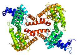
Human GC is a glycosylated alpha-globulin, ~58 kDa in size. Its 458 amino acids are coded for by 1690 nucleotides on chromosome 4 (4q11–q13). The primary structure contains 28 cysteine residues forming multiple disulfide bonds. GC contains 3 domains. Domain 1 is composed of 10 alpha helices, domain 2 of 9, and domain 3 of 4.[8]
It is able to bind the various forms of vitamin D including ergocalciferol (vitamin D2) and cholecalciferol (vitamin D3), the 25-hydroxylated forms (calcifediol), and the active hormonal product, 1,25-dihydroxyvitamin D (calcitriol). The major proportion of vitamin D in blood is bound to this protein. It transports vitamin D metabolites between skin, liver and kidney, and then on to the various target tissues.[6][9]
Beyond acting as the carrier protein for vitamin D and its metabolites, DBP also transports free fatty acids,[10] binds to actin[11] and may help prevent actin polymerization during tissue injury.[12] It also might serve as a macrophage activator, contributing to the inflammatory response by modulating T-cell activity.[13]
The transcription factors HFN1α is a positive regulator while HFN1β is a dominant negative regulator of DBP expression.[15]
Evolution
Phylogenetic analyses suggest that DBP diverged from ancestral albumin through gene duplication events that occurred after the separation of jawless fish (cyclostomes) from jawed vertebrates approximately 450 million years ago.[16] This timeline is supported by the apparent absence of DBP-like proteins in lampreys and hagfish, though these organisms retain vitamin D transport capability through alternative lipoprotein-mediated mechanisms.[17] DBP is found throughout jawed vertebrates, from bony fish to mammals, suggesting its evolution coincided with the development of calcified skeletons and more sophisticated calcium homeostasis requirements.[18]
Variation
Many genetic variants of the GC gene are known. They produce 6 main haplotypes and 3 main protein variants (Gc1S, Gc1F and Gc2).[19] The genetic variations are associated with differences in circulating 25-hydroxyvitamin D levels.[20] They have been proposed to account for some of the differences in vitamin D status in different ethnic groups,[21] and have been found to correlate with the response to vitamin D supplementation.[19]
↑"Human PubMed Reference:". National Center for Biotechnology Information, U.S. National Library of Medicine.
↑"Mouse PubMed Reference:". National Center for Biotechnology Information, U.S. National Library of Medicine.
↑Mikkelsen M, Jacobsen P, Henningsen K (Jul 1977). "Possible localization of Gc-System on chromosome 4. Loss of long arm 4 material associated with father-child incompatibility within the Gc-System". Human Heredity. 27 (2): 105–107. doi:10.1159/000152857. PMID558959.
↑Verboven C, Rabijns A, De Maeyer M, Van Baelen H, Bouillon R, De Ranter C (February 2002). "A structural basis for the unique binding features of the human vitamin D-binding protein". Nature Structural Biology. 9 (2): 131–136. doi:10.1038/nsb754. PMID11799400. S2CID38990672.
↑Williams MH, Van Alstyne EL, Galbraith RM (June 1988). "Evidence of a novel association of unsaturated fatty acids with Gc (vitamin D-binding protein)". Biochemical and Biophysical Research Communications. 153 (3): 1019–1024. doi:10.1016/S0006-291X(88)81330-5. PMID3134016.
↑Meier U, Gressner O, Lammert F, Gressner AM (July 2006). "Gc-globulin: roles in response to injury". Clinical Chemistry. 52 (7): 1247–1253. doi:10.1373/clinchem.2005.065680. PMID16709624.
↑Delanghe JR, Speeckaert R, Speeckaert MM (October 2015). "Behind the scenes of vitamin D binding protein: more than vitamin D binding". Best Practice & Research. Clinical Endocrinology & Metabolism. 29 (5): 773–786. doi:10.1016/j.beem.2015.06.006. PMID26522461.
↑Hay AW, Watson G (1976). "The plasma transport proteins of 25-hydroxycholecalciferol in fish, amphibians, reptiles and birds". Comparative Biochemistry and Physiology. B, Comparative Biochemistry. 53 (2): 167–172. doi:10.1016/0305-0491(76)90029-8. PMID1253553.
↑McGrath JJ, Saha S, Burne TH, Eyles DW (July 2010). "A systematic review of the association between common single nucleotide polymorphisms and 25-hydroxyvitamin D concentrations". The Journal of Steroid Biochemistry and Molecular Biology. 121 (1–2): 471–477. doi:10.1016/j.jsbmb.2010.03.073. PMID20363324. S2CID20057294.
Svasti J, Kurosky A, Bennett A, Bowman BH (April 1979). "Molecular basis for the three major forms of human serum vitamin D binding protein (group-specific component)". Biochemistry. 18 (8): 1611–1617. doi:10.1021/bi00575a036. PMID218624.
Braun A, Bichlmaier R, Cleve H (June 1992). "Molecular analysis of the gene for the human vitamin-D-binding protein (group-specific component): allelic differences of the common genetic GC types". Human Genetics. 89 (4): 401–406. doi:10.1007/BF00194311. PMID1352271. S2CID1913655.
Szpirer C, Riviere M, Cortese R, Nakamura T, Islam MQ, Levan G, etal. (June 1992). "Chromosomal localization in man and rat of the genes encoding the liver-enriched transcription factors C/EBP, DBP, and HNF1/LFB-1 (CEBP, DBP, and transcription factor 1, TCF1, respectively) and of the hepatocyte growth factor/scatter factor gene (HGF)". Genomics. 13 (2): 293–300. doi:10.1016/0888-7543(92)90245-N. PMID1535333.
Dawson SJ, White LA (May 1992). "Treatment of Haemophilus aphrophilus endocarditis with ciprofloxacin". The Journal of Infection. 24 (3): 317–320. doi:10.1016/S0163-4453(05)80037-4. PMID1602151.
Yang F, Bergeron JM, Linehan LA, Lalley PA, Sakaguchi AY, Bowman BH (August 1990). "Mapping and conservation of the group-specific component gene in mouse". Genomics. 7 (4): 509–516. doi:10.1016/0888-7543(90)90193-X. PMID1696927.
Schoentgen F, Metz-Boutigue MH, Jollès J, Constans J, Jollès P (June 1986). "Complete amino acid sequence of human vitamin D-binding protein (group-specific component): evidence of a three-fold internal homology as in serum albumin and alpha-fetoprotein". Biochimica et Biophysica Acta (BBA) - Protein Structure and Molecular Enzymology. 871 (2): 189–198. doi:10.1016/0167-4838(86)90173-1. PMID2423133.
Yang F, Naberhaus KH, Adrian GS, Gardella JM, Brissenden JE, Bowman BH (1987). "The vitamin D-binding protein gene contains conserved nucleotide sequences that respond to heavy metal, adipocyte and mitotic signals". Gene. 54 (2–3): 285–290. doi:10.1016/0378-1119(87)90499-9. PMID2958390.
Cooke NE, Willard HF, David EV, George DL (July 1986). "Direct regional assignment of the gene for vitamin D binding protein (Gc-globulin) to human chromosome 4q11-q13 and identification of an associated DNA polymorphism". Human Genetics. 73 (3): 225–229. doi:10.1007/BF00401232. PMID3015768. S2CID38816588.
Nestler JE, McLeod JF, Kowalski MA, Strauss JF, Haddad JG (May 1987). "Detection of vitamin D binding protein on the surface of cytotrophoblasts isolated from human placentae". Endocrinology. 120 (5): 1996–2002. doi:10.1210/endo-120-5-1996. PMID3552627.
Constans J, Oksman F, Viau M (August 1981). "Binding of the apo and holo forms of the serum vitamin D-binding protein to human lymphocyte cytoplasm and membrane by indirect immunofluorescence". Immunology Letters. 3 (3): 159–162. doi:10.1016/0165-2478(81)90120-6. PMID7026425.
Braun A, Kofler A, Morawietz S, Cleve H (December 1993). "Sequence and organization of the human vitamin D-binding protein gene". Biochimica et Biophysica Acta (BBA) - Gene Structure and Expression. 1216 (3): 385–394. doi:10.1016/0167-4781(93)90005-x. PMID7505619.
1j78: Crystallographic analysis of the human vitamin D binding protein
1j7e: A Structural Basis for the Unique Binding Features of the Human Vitamin D-binding Protein
1kw2: CRYSTAL STRUCTURE OF UNCOMPLEXED VITAMIN D-BINDING PROTEIN
1kxp: CRYSTAL STRUCTURE OF HUMAN VITAMIN D-BINDING PROTEIN IN COMPLEX WITH SKELETAL ACTIN
1lot: CRYSTAL STRUCTURE OF THE COMPLEX OF ACTIN WITH VITAMIN D-BINDING PROTEIN
1ma9: Crystal structure of the complex of human vitamin D binding protein and rabbit muscle actin
This page is based on this Wikipedia article Text is available under the CC BY-SA 4.0 license; additional terms may apply. Images, videos and audio are available under their respective licenses.