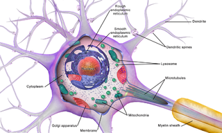
Interneuron

Last updated
Interneuron
Interneuron566-01.svg
Cartoon of a locust interneuron that integrates information about wind in order to control wing motor neurons during flight [1]
Details
Location Nervous system
Identifiers
MeSH D007395
NeuroLex ID birnlex_2534
TH H2.00.06.1.00058
FMA 67313
Anatomical terms of neuroanatomy

Interneurons (also called internuncial neurons, relay neurons, association neurons, connector neurons, intermediate neurons or local circuit neurons) are neurons that connect to brain regions, i.e. not direct motor neurons or sensory neurons. Interneurons are the central nodes of neural circuits, enabling communication between sensory or motor neurons and the central nervous system (CNS). [2] They play vital roles in reflexes, neuronal oscillations, [3] and neurogenesis in the adult mammalian brain.[ citation needed ]

Contents

Interneurons can be further broken down into two groups: local interneurons and relay interneurons. [4] [ need quotation to verify ] Local interneurons have short axons and form circuits with nearby neurons to analyze small pieces of information. [5] Relay interneurons have long axons and connect circuits of neurons in one region of the brain with those in other regions. [5] However, interneurons are generally considered to operate mainly within local brain areas. [6] The interaction between interneurons allow the brain to perform complex functions such as learning, and decision-making.

Structure

Approximately 20–30% of the neurons in the neocortex are interneurons, while the remaining neurons are pyramidal neurons. [7] Investigations into the molecular diversity of neurons is impeded by the inability to isolate cell populations born at different times for gene expression analysis. An effective means of identifying coetaneous interneurons is neuronal birthdating. [8] This can be achieved using nucleoside analogs such as EdU. [9] [8]

In 2008, a nomenclature for the features of GABAergic cortical interneurons was proposed, called Petilla terminology. [10]

Spinal cord

Cortex

Cerebellum

Striatum

Function

Interneurons in the CNS are primarily inhibitory, and use the neurotransmitter GABA or glycine. However, excitatory interneurons using glutamate in the CNS also exist, as do interneurons releasing neuromodulators like acetylcholine.

In addition to these general functions, interneurons in the insect CNS play a number of specific roles in different parts of the nervous system, and also are either excitatory or inhibitory. For example, in the olfactory system, interneurons are responsible for integrating information from odorant receptors and sending signals to the mushroom bodies, which are involved in learning and memory. [17] [18] In the visual system, interneurons are responsible for processing motion information and sending signals to the optic lobes, which are involved in visual navigation. [19] [20]

Interneurons are also important for coordinating complex behaviors, such as flight and locomotion. For example, interneurons in the thoracic ganglia are responsible for coordinating the activity of the leg muscles during walking [21] and flying. [22]

Interneurons main function is to provide a neural circuit, conducting flow of signals or information between a sensory neuron and or motor neuron. [23]

See also

Related Research Articles

<span class="mw-page-title-main">Neuron</span> Electrically excitable cell found in the nervous system of animals

Within a nervous system, a neuron, neurone, or nerve cell is an electrically excitable cell that fires electric signals called action potentials across a neural network. Neurons communicate with other cells via synapses, which are specialized connections that commonly use minute amounts of chemical neurotransmitters to pass the electric signal from the presynaptic neuron to the target cell through the synaptic gap.

<span class="mw-page-title-main">Striatum</span> Nucleus in the basal ganglia of the brain

The striatum, or corpus striatum, is a nucleus in the subcortical basal ganglia of the forebrain. The striatum is a critical component of the motor and reward systems; receives glutamatergic and dopaminergic inputs from different sources; and serves as the primary input to the rest of the basal ganglia.

The development of the nervous system, or neural development (neurodevelopment), refers to the processes that generate, shape, and reshape the nervous system of animals, from the earliest stages of embryonic development to adulthood. The field of neural development draws on both neuroscience and developmental biology to describe and provide insight into the cellular and molecular mechanisms by which complex nervous systems develop, from nematodes and fruit flies to mammals.

<span class="mw-page-title-main">Olfactory bulb</span> Neural structure

The olfactory bulb is a neural structure of the vertebrate forebrain involved in olfaction, the sense of smell. It sends olfactory information to be further processed in the amygdala, the orbitofrontal cortex (OFC) and the hippocampus where it plays a role in emotion, memory and learning. The bulb is divided into two distinct structures: the main olfactory bulb and the accessory olfactory bulb. The main olfactory bulb connects to the amygdala via the piriform cortex of the primary olfactory cortex and directly projects from the main olfactory bulb to specific amygdala areas. The accessory olfactory bulb resides on the dorsal-posterior region of the main olfactory bulb and forms a parallel pathway. Destruction of the olfactory bulb results in ipsilateral anosmia, while irritative lesions of the uncus can result in olfactory and gustatory hallucinations.

<span class="mw-page-title-main">Nigrostriatal pathway</span> Bilateral pathway in the brain

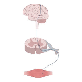
The nigrostriatal pathway is a bilateral dopaminergic pathway in the brain that connects the substantia nigra pars compacta (SNc) in the midbrain with the dorsal striatum in the forebrain. It is one of the four major dopamine pathways in the brain, and is critical in the production of movement as part of a system called the basal ganglia motor loop. Dopaminergic neurons of this pathway release dopamine from axon terminals that synapse onto GABAergic medium spiny neurons (MSNs), also known as spiny projection neurons (SPNs), located in the striatum.

<span class="mw-page-title-main">Ventral tegmental area</span> Group of neurons on the floor of the midbrain

The ventral tegmental area (VTA), also known as the ventral tegmental area of Tsai, or simply ventral tegmentum, is a group of neurons located close to the midline on the floor of the midbrain. The VTA is the origin of the dopaminergic cell bodies of the mesocorticolimbic dopamine system and other dopamine pathways; it is widely implicated in the drug and natural reward circuitry of the brain. The VTA plays an important role in a number of processes, including reward cognition and orgasm, among others, as well as several psychiatric disorders. Neurons in the VTA project to numerous areas of the brain, ranging from the prefrontal cortex to the caudal brainstem and several regions in between.

<span class="mw-page-title-main">Basket cell</span>

Basket cells are inhibitory GABAergic interneurons of the brain, found throughout different regions of the cortex and cerebellum.

<span class="mw-page-title-main">Rostral migratory stream</span> One path neural stem cells take to reach the olfactory bulb


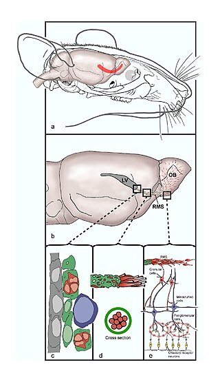
The rostral migratory stream (RMS) is a specialized migratory route found in the brain of some animals along which neuronal precursors that originated in the subventricular zone (SVZ) of the brain migrate to reach the main olfactory bulb (OB). The importance of the RMS lies in its ability to refine and even change an animal's sensitivity to smells, which explains its importance and larger size in the rodent brain as compared to the human brain, as our olfactory sense is not as developed. This pathway has been studied in the rodent, rabbit, and both the squirrel monkey and rhesus monkey. When the neurons reach the OB they differentiate into GABAergic interneurons as they are integrated into either the granule cell layer or periglomerular layer.

The antennal lobe is the primary olfactory brain area in insects. The antennal lobe is a sphere-shaped deutocerebral neuropil in the brain that receives input from the olfactory sensory neurons in the antennae and mouthparts. Functionally, it shares some similarities with the olfactory bulb in vertebrates. The anatomy and physiology function of the insect brain can be studied by dissecting open the insect brain and imaging or carrying out in vivo electrophysiological recordings from it.

Neuromodulation is the physiological process by which a given neuron uses one or more chemicals to regulate diverse populations of neurons. Neuromodulators typically bind to metabotropic, G-protein coupled receptors (GPCRs) to initiate a second messenger signaling cascade that induces a broad, long-lasting signal. This modulation can last for hundreds of milliseconds to several minutes. Some of the effects of neuromodulators include: altering intrinsic firing activity, increasing or decreasing voltage-dependent currents, altering synaptic efficacy, increasing bursting activity and reconfigurating synaptic connectivity.

<span class="mw-page-title-main">Medium spiny neuron</span> Type of GABAergic neuron in the striatum

Medium spiny neurons (MSNs), also known as spiny projection neurons (SPNs), are a special type of GABAergic inhibitory cell representing 95% of neurons within the human striatum, a basal ganglia structure. Medium spiny neurons have two primary phenotypes : D1-type MSNs of the direct pathway and D2-type MSNs of the indirect pathway. Most striatal MSNs contain only D1-type or D2-type dopamine receptors, but a subpopulation of MSNs exhibit both phenotypes.

<span class="mw-page-title-main">Mossy fiber (hippocampus)</span> Pathway in the hippocampus

In the hippocampus, the mossy fiber pathway consists of unmyelinated axons projecting from granule cells in the dentate gyrus that terminate on modulatory hilar mossy cells and in Cornu Ammonis area 3 (CA3), a region involved in encoding short-term memory. These axons were first described as mossy fibers by Santiago Ramón y Cajal as they displayed varicosities along their lengths that gave them a mossy appearance. The axons that make up the pathway emerge from the basal portions of the granule cells and pass through the hilus of the dentate gyrus before entering the stratum lucidum of CA3. Granule cell synapses tend to be glutamatergic, though immunohistological data has indicated that some synapses contain neuropeptidergic elements including opiate peptides such as dynorphin and enkephalin. There is also evidence for co-localization of both GABAergic and glutamatergic neurotransmitters within mossy fiber terminals. GABAergic and glutamatergic co-localization in mossy fiber boutons has been observed primarily in the developing hippocampus, but in adulthood, evidence suggests that mossy fiber synapses may alternate which neurotransmitter is released through activity-dependent regulation.

Optogenetics is a biological technique to control the activity of neurons or other cell types with light. This is achieved by expression of light-sensitive ion channels, pumps or enzymes specifically in the target cells. On the level of individual cells, light-activated enzymes and transcription factors allow precise control of biochemical signaling pathways. In systems neuroscience, the ability to control the activity of a genetically defined set of neurons has been used to understand their contribution to decision making, learning, fear memory, mating, addiction, feeding, and locomotion. In a first medical application of optogenetic technology, vision was partially restored in a blind patient.

Mosaic analysis with a repressible cell marker, or MARCM, is a genetics technique for creating individually labeled homozygous cells in an otherwise heterozygous Drosophila melanogaster. It has been a crucial tool in studying the development of the Drosophila nervous system. This technique relies on recombination during mitosis mediated by FLP-FRT recombination. As one copy of a gene, provided by the balancer chromosome, is often enough to rescue a mutant phenotype, MARCM clones can be used to study a mutant phenotype in an otherwise wildtype animal.

Sharp waves and ripples (SWRs) are oscillatory patterns produced by extremely synchronised activity of neurons in the mammalian hippocampus and neighbouring regions which occur spontaneously in idle waking states or during NREM sleep. They can be observed with a variety of imaging methods, such as EEG. They are composed of large amplitude sharp waves in local field potential and produced by tens of thousands of neurons firing together within 30–100 ms window. They are some of the most synchronous oscillations patterns in the brain, making them susceptible to pathological patterns such as epilepsy.They have been extensively characterised and described by György Buzsáki and have been shown to be involved in memory consolidation in NREM sleep and the replay of memories acquired during wakefulness.

<span class="mw-page-title-main">Neuronal lineage marker</span> Endogenous tag expressed in different cells along neurogenesis and differentiated cells

A neuronal lineage marker is an endogenous tag that is expressed in different cells along neurogenesis and differentiated cells such as neurons. It allows detection and identification of cells by using different techniques. A neuronal lineage marker can be either DNA, mRNA or RNA expressed in a cell of interest. It can also be a protein tag, as a partial protein, a protein or an epitope that discriminates between different cell types or different states of a common cell. An ideal marker is specific to a given cell type in normal conditions and/or during injury. Cell markers are very valuable tools for examining the function of cells in normal conditions as well as during disease. The discovery of various proteins specific to certain cells led to the production of cell-type-specific antibodies that have been used to identify cells.

Neurogenesis is the process by which nervous system cells, the neurons, are produced by neural stem cells (NSCs). In short, it is brain growth in relation to its organization. This occurs in all species of animals except the porifera (sponges) and placozoans. Types of NSCs include neuroepithelial cells (NECs), radial glial cells (RGCs), basal progenitors (BPs), intermediate neuronal precursors (INPs), subventricular zone astrocytes, and subgranular zone radial astrocytes, among others.

Neurogliaform cells (NGF) are inhibitory (GABAergic) interneurons found in the cortex and the hippocampus. NGF cells represent approximately 10% of the total hippocampal inhibitory interneuron population.

An axo-axonic synapse is a type of synapse, formed by one neuron projecting its axon terminals onto another neuron's axon.

<span class="mw-page-title-main">Brain cell</span> Functional tissue of the brain

Brain cells make up the functional tissue of the brain. The rest of the brain tissue is structural or connective called the stroma which includes blood vessels. The two main types of cells in the brain are neurons, also known as nerve cells, and glial cells, also known as neuroglia.

References

  1. Pearson, K. G. and Wolf, H. Connections of hindwing tegulae with flight neurones in the locust, Locusta migratoria. J. Exp. Biol. 135: 381-409, 1988
  2. "Types of neurons - Queensland Brain Institute - University of Queensland". 9 November 2017.
  3. Whittington, M.A; Traub, R.D; Kopell, N; Ermentrout, B; Buhl, E.H (2000). "Inhibition-based rhythms: Experimental and mathematical observations on network dynamics". International Journal of Psychophysiology. 38 (3): 315–36. CiteSeerX   10.1.1.16.6410 . doi:10.1016/S0167-8760(00)00173-2. PMID   11102670.
  4. Carlson, Neil R. (2013). Physiology of Behavior (11th ed.). Pearson Higher Education. p.  28. ISBN   978-0-205-23939-9.
  5. 1 2 Kandel, Eric; Schwartz, James; Jessell, Thomas, eds. (2000). Principles of Neural Science (4th ed.). New York City, New York: McGraw Hill Companies. p.  25. ISBN   978-0-8385-7701-1.
  6. Kepecs, Adam; Fishell, Gordon (2014). "Interneuron Cell Types: Fit to form and formed to fit". Nature. Nature , 2014 HHS Public Access pp 10, 28. 505 (7483): 318–326. doi:10.1038/nature12983. PMC   4349583 . PMID   24429630.
  7. Markram, Henry; et al. (2004). "Interneurons of the neocortical inhibitory system". Nature Reviews Neuroscience. 5 (10): 793–807. doi:10.1038/nrn1519. PMID   15378039. S2CID   382334.
  8. 1 2 Ng, Hui Xuan; Lee, Ean Phing; Cavanagh, Brenton L.; Britto, Joanne M.; Tan, Seong-Seng (2017). "A method for isolating cortical interneurons sharing the same birthdays for gene expression studies". Experimental Neurology. 295: 36–45. doi:10.1016/j.expneurol.2017.05.006. PMID   28511841. S2CID   3377296.
  9. Endaya, Berwini; Cavanagh, Brenton; Alowaidi, Faisal; Walker, Tom; Pennington, Nicholas de; Ng, Jin-Ming A.; Lam, Paula Y.P.; Mackay-Sim, Alan; Neuzil, Jiri (2016). "Isolating dividing neural and brain tumour cells for gene expression profiling". Journal of Neuroscience Methods. 257: 121–133. doi:10.1016/j.jneumeth.2015.09.020. PMID   26432933. S2CID   44969376.
  10. Ascoli, Giorgio A.; Alonso-Nanclares, Lidia; Anderson, Stewart A.; Barrionuevo, German; Benavides-Piccione, Ruth; Burkhalter, Andreas; Buzsáki, György; Cauli, Bruno; Defelipe, Javier; Fairén, Alfonso; Feldmeyer, Dirk; Fishell, Gord; Fregnac, Yves; Freund, Tamas F.; Gardner, Daniel; Gardner, Esther P.; Goldberg, Jesse H.; Helmstaedter, Moritz; Hestrin, Shaul; Karube, Fuyuki; Kisvárday, Zoltán F.; Lambolez, Bertrand; Lewis, David A.; Marin, Oscar; Markram, Henry; Muñoz, Alberto; Packer, Adam; Petersen, Carl C. H.; Rockland, Kathleen S.; et al. (2008). "Petilla terminology: Nomenclature of features of GABAergic interneurons of the cerebral cortex". Nature Reviews Neuroscience. 9 (7): 557–68. doi:10.1038/nrn2402. PMC   2868386 . PMID   18568015.
  11. Muñoz, W; Tremblay, R; Levenstein, D; Rudy, B (3 March 2017). "Layer-specific modulation of neocortical dendritic inhibition during active wakefulness". Science. 355 (6328): 954–959. Bibcode:2017Sci...355..954M. doi: 10.1126/science.aag2599 . PMID   28254942.
  12. Tepper, James M.; Koós, Tibor (1999). "Inhibitory control of neostriatal projection neurons by GABAergic interneurons". Nature Neuroscience. 2 (5): 467–72. doi:10.1038/8138. PMID   10321252. S2CID   16088859.
  13. Zhou, Fu-Ming; Wilson, Charles J.; Dani, John A. (2002). "Cholinergic interneuron characteristics and nicotinic properties in the striatum". Journal of Neurobiology. 53 (4): 590–605. doi: 10.1002/neu.10150 . PMID   12436423.
  14. English, Daniel F; Ibanez-Sandoval, Osvaldo; Stark, Eran; Tecuapetla, Fatuel; Buzsáki, György; Deisseroth, Karl; Tepper, James M; Koos, Tibor (2011). "GABAergic circuits mediate the reinforcement-related signals of striatal cholinergic interneurons". Nature Neuroscience. 15 (1): 123–30. doi:10.1038/nn.2984. PMC   3245803 . PMID   22158514.
  15. Ibanez-Sandoval, O.; Tecuapetla, F.; Unal, B.; Shah, F.; Koos, T.; Tepper, J. M. (2010). "Electrophysiological and Morphological Characteristics and Synaptic Connectivity of Tyrosine Hydroxylase-Expressing Neurons in Adult Mouse Striatum". Journal of Neuroscience. 30 (20): 6999–7016. doi:10.1523/JNEUROSCI.5996-09.2010. PMC   4447206 . PMID   20484642.
  16. 1 2 Ibáñez-Sandoval, Osvaldo; Koós, Tibor; Tecuapetla, Fatuel; Tepper, James M. (2010). "Heterogeneity and Diversity of Striatal GABAergic Interneurons". Frontiers in Neuroanatomy. 4: 150. doi: 10.3389/fnana.2010.00150 . PMC   3016690 . PMID   21228905.
  17. Liou, Nan-Fu; Lin, Shih-Han; Chen, Ying-Jun; Tsai, Kuo-Ting; Yang, Chi-Jen; Lin, Tzi-Yang; Wu, Ting-Han; Lin, Hsin-Ju; Chen, Yuh-Tarng; Gohl, Daryl M.; Silies, Marion; Chou, Ya-Hui (2018-06-08). "Diverse populations of local interneurons integrate into the Drosophila adult olfactory circuit". Nature Communications. 9 (1): 2232. Bibcode:2018NatCo...9.2232L. doi:10.1038/s41467-018-04675-x. ISSN   2041-1723. PMC   5993751 . PMID   29884811.
  18. Zheng, Zhihao; Li, Feng; Fisher, Corey; Ali, Iqbal J.; Sharifi, Nadiya; Calle-Schuler, Steven; Hsu, Joseph; Masoodpanah, Najla; Kmecova, Lucia; Kazimiers, Tom; Perlman, Eric; Nichols, Matthew; Li, Peter H.; Jain, Viren; Bock, Davi D. (August 2022). "Structured sampling of olfactory input by the fly mushroom body". Current Biology. 32 (15): 3334–3349.e6. doi:10.1016/j.cub.2022.06.031. ISSN   0960-9822. PMC   9413950 . PMID   35797998.
  19. Zhu, Yan (2013-07-29). "The Drosophila visual system: From neural circuits to behavior". Cell Adhesion & Migration. 7 (4): 333–344. doi:10.4161/cam.25521. ISSN   1933-6918. PMC   3739809 . PMID   23880926.
  20. Shinomiya, Kazunori; Nern, Aljoscha; Meinertzhagen, Ian A.; Plaza, Stephen M.; Reiser, Michael B. (August 2022). "Neuronal circuits integrating visual motion information in Drosophila melanogaster". Current Biology. 32 (16): 3529–3544.e2. doi: 10.1016/j.cub.2022.06.061 . ISSN   0960-9822.
  21. Bidaye, Salil S.; Laturney, Meghan; Chang, Amy K.; Liu, Yuejiang; Bockemühl, Till; Büschges, Ansgar; Scott, Kristin (November 2020). "Two Brain Pathways Initiate Distinct Forward Walking Programs in Drosophila". Neuron. 108 (3): 469–485.e8. doi:10.1016/j.neuron.2020.07.032. PMC   9435592 . PMID   32822613.
  22. King, David G.; Wyman, Robert J. (1980-12-01). "Anatomy of the giant fibre pathway inDrosophila. I. Three thoracic components of the pathway". Journal of Neurocytology. 9 (6): 753–770. doi:10.1007/BF01205017. ISSN   1573-7381. PMID   6782199. S2CID   10530883.
  23. "Types of Neurons". University of Queensland. Queensland Brain Institute. 9 November 2017. Retrieved 26 April 2023.