
The brainstem is the posterior stalk-like part of the brain that connects the cerebrum with the spinal cord. In the human brain the brainstem is composed of the midbrain, the pons, and the medulla oblongata. The midbrain is continuous with the thalamus of the diencephalon through the tentorial notch, and sometimes the diencephalon is included in the brainstem.

The pyramidal tracts include both the corticobulbar tract and the corticospinal tract. These are aggregations of efferent nerve fibers from the upper motor neurons that travel from the cerebral cortex and terminate either in the brainstem (corticobulbar) or spinal cord (corticospinal) and are involved in the control of motor functions of the body.

The solitary nucleus(SN) (nucleus of the solitary tract, nucleus solitarius, or nucleus tractus solitarii) is a series of neurons whose cell bodies form a roughly vertical column of grey matter in the medulla oblongata of the brainstem. Their axons form the bulk of the enclosed solitary tract. The solitary nucleus can be divided into different parts including dorsomedial, dorsolateral, and ventrolateral subnuclei.

The medial longitudinal fasciculus (MLF) is a prominent bundle of nerve fibres which pass within the ventral/anterior portion of periaqueductal gray of the mesencephalon (midbrain). It contains the interstitial nucleus of Cajal, responsible for oculomotor control, head posture, and vertical eye movement.

The reticular formation is a set of interconnected nuclei in the brainstem that spans from the lower end of the medulla oblongata to the upper end of the midbrain. The neurons of the reticular formation make up a complex set of neural networks in the core of the brainstem. The reticular formation is made up of a diffuse net-like formation of reticular nuclei which is not well-defined. It may be seen as being made up of all the interspersed cells in the brainstem between the more compact and named structures.

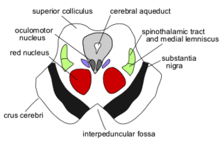
The red nucleus or nucleus ruber is a structure in the rostral midbrain involved in motor coordination. The red nucleus is pale pink, which is believed to be due to the presence of iron in at least two different forms: hemoglobin and ferritin. The structure is located in the midbrain tegmentum next to the substantia nigra and comprises caudal magnocellular and rostral parvocellular components. The red nucleus and substantia nigra are subcortical centers of the extrapyramidal motor system.

The spinocerebellar tracts are nerve tracts originating in the spinal cord and terminating in the same side (ipsilateral) of the cerebellum. The two main tracts are the dorsal spinocerebellar tract, and the ventral spinocerebellar tract. Both of these tracts are located in the peripheral region of the lateral funiculi. Other tracts are the rostral spinocerebellar tract, and the cuneocerebellar tract.

The globose nucleus is one of the deep cerebellar nuclei. It is located medial to the emboliform nucleus, and lateral to the fastigial nucleus. The globose nucleus and emboliform nucleus are known collectively as the interposed nuclei.
The interposed nucleus is the combined paired globose and emboliform nuclei, on either side of the cerebellum. It is located in the roof of the fourth ventricle, lateral to the fastigial nucleus. The emboliform nucleus is the anterior interposed nucleus, and the globose nucleus is the posterior interposed nucleus.

The accessory cuneate nucleus is a nucleus situated in the caudal medulla oblongata just lateral to the cuneate nucleus. It relays unconscious proprioceptive sensory information from the upper limb and upper trunk to the cerebellum via the cuneocerebellar fibers.

The vestibulospinal tract is a nerve tract in the central nervous system. Specifically, it is a component of the extrapyramidal system and is classified as a component of the medial pathway. Like other descending motor pathways, the vestibulospinal fibers of the tract relay information from nuclei to motor neurons. The vestibular nuclei receive information through the vestibulocochlear nerve about changes in the orientation of the head. The nuclei relay motor commands through the vestibulospinal tract. The function of these motor commands is to alter muscle tone, extend, and change the position of the limbs and head with the goal of supporting posture and maintaining balance of the body and head.
The nucleus raphe magnus (NRM) is one of the seven raphe nuclei. It is situated in the pons in the brainstem, just rostral to the nucleus raphe obscurus.

The Rexed laminae comprise a system of ten layers of grey matter (I–X), identified in the early 1950s by Bror Rexed to label portions of the grey columns of the spinal cord.

Alpha (α) motor neurons (also called alpha motoneurons), are large, multipolar lower motor neurons of the brainstem and spinal cord. They innervate extrafusal muscle fibers of skeletal muscle and are directly responsible for initiating their contraction. Alpha motor neurons are distinct from gamma motor neurons, which innervate intrafusal muscle fibers of muscle spindles.
The gigantocellular reticular nucleus is the (efferent/motor) medial zone of the reticular formation of the caudal pons and rostral medulla oblongata. It consists of a substantial number of giant neurons, but also contains small and medium sized neurons.

The basilar part of pons, also known as basis pontis, or basilar pons, is the ventral part of the pons in the brainstem; the dorsal part is known as the pontine tegmentum.
The central tegmental tract is a tract that carries ascending and descending fibers, situated in the midbrain tegmentum, and the pontine tegmentum. The tract is situated in the central portion of the reticular formation.
Corticopontine fibers are projections from layer V of the cerebral cortex to the pontine nuclei of the ventral pons. They represent the first link in a cortico-cerebello-cortical pathway mediating neocerebellar control of the motor cortex. The pathway is especially important for voluntary movements.
The hypothalamospinal tract is an unmyelinated non-decussated descending nerve tract that arises in the hypothalamus and projects to the brainstem and spinal cord to synapse with pre-ganglionic autonomic neurons.

The spinomesencephalic pathway, spinomesencephalic tract or spino-quadrigeminal system of Mott, includes a number of ascending tracts in the spinal cord, including the spinotectal tract. The spinomesencephalic tract is one of the ascending tracts in the anterolateral system of the spinal cord that projects to various parts of the midbrain. It is involved in the processing of pain and visceral sensations.

