Red nucleus

Last updated
Red nucleus
Cn3nucleus-en.svg
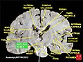
Transverse section through the midbrain showing the location of the red nuclei. The superior colliculi are at the top of image and the cerebral peduncles at the bottom of image – both in section.
Details
Part of Midbrain
Identifiers
Latin nucleus ruber
MeSH D012012
NeuroNames 505
NeuroLex ID birnlex_1478
TA98 A14.1.06.323
TA2 5898
FMA 62407
Anatomical terms of neuroanatomy

The red nucleus or nucleus ruber is a structure in the rostral midbrain involved in motor coordination. [1] The red nucleus is pale pink, which is believed to be due to the presence of iron in at least two different forms: hemoglobin and ferritin. [2] [3] The structure is located in the tegmentum of the midbrain next to the substantia nigra and comprises caudal magnocellular and rostral parvocellular components. [1] The red nucleus and substantia nigra are subcortical centers of the extrapyramidal motor system.

Contents

Function

In a vertebrate without a significant corticospinal tract, gait is mainly controlled by the red nucleus. [4] However, in primates, where the corticospinal tract is dominant, the rubrospinal tract may be regarded as vestigial in motor function. Therefore, the red nucleus is less important in primates than in many other mammals. [1] [5] Nevertheless, the crawling of babies is controlled by the red nucleus, as is arm swinging in typical walking. [6] The red nucleus may play an additional role in controlling muscles of the shoulder and upper arm via projections of its magnocellular part. [7] [8] In humans, the red nucleus also has limited control over hands, as the rubrospinal tract is more involved in large muscle movement such as that for the arms (but not for the legs, as the tract terminates in the superior thoracic region of the spinal cord). Fine control of the fingers is not modified by the functioning of the red nucleus but relies on the corticospinal tract. [9] The majority of red nucleus axons do not project to the spinal cord but, via its parvocellular part, relay information from the motor cortex to the cerebellum through the inferior olivary complex, an important relay center in the medulla. [1]

Input and output

The red nucleus receives many inputs from the cerebellum (interposed nucleus and the lateral cerebellar nucleus) of the opposite side and an input from the motor cortex of the same side. [10]

The red nucleus has two sets of efferents: [10]

See also

Additional images

Related Research Articles

<span class="mw-page-title-main">Brainstem</span> Posterior part of the brain, adjoining and structurally continuous

The brainstem is the stalk-like part of the brain that interconnects the cerebrum and diencephalon with the spinal cord. In the human brain, the brainstem is composed of the midbrain, the pons, and the medulla oblongata. The midbrain is continuous with the thalamus of the diencephalon through the tentorial notch.

<span class="mw-page-title-main">Extrapyramidal system</span> Connection between brain and spinal cord

In anatomy, the extrapyramidal system is a part of the motor system network causing involuntary actions. The system is called extrapyramidal to distinguish it from the tracts of the motor cortex that reach their targets by traveling through the pyramids of the medulla. The pyramidal tracts may directly innervate motor neurons of the spinal cord or brainstem, whereas the extrapyramidal system centers on the modulation and regulation of anterior (ventral) horn cells.

<span class="mw-page-title-main">Internal capsule</span> White matter structure situated in the inferomedial part of each cerebral hemisphere of the brain

The internal capsule is a white matter structure situated in the inferomedial part of each cerebral hemisphere of the brain. It carries information past the basal ganglia, separating the caudate nucleus and the thalamus from the putamen and the globus pallidus. The internal capsule contains both ascending and descending axons, going to and coming from the cerebral cortex. It also separates the caudate nucleus and the putamen in the dorsal striatum, a brain region involved in motor and reward pathways.

<span class="mw-page-title-main">Neural pathway</span> Connection formed between neurons that allows neurotransmission

In neuroanatomy, a neural pathway is the connection formed by axons that project from neurons to make synapses onto neurons in another location, to enable neurotransmission. Neurons are connected by a single axon, or by a bundle of axons known as a nerve tract, or fasciculus. Shorter neural pathways are found within grey matter in the brain, whereas longer projections, made up of myelinated axons, constitute white matter.

<span class="mw-page-title-main">Pyramidal tracts</span> The corticobulbar tract and the corticospinal tract

The pyramidal tracts include both the corticobulbar tract and the corticospinal tract. These are aggregations of efferent nerve fibers from the upper motor neurons that travel from the cerebral cortex and terminate either in the brainstem (corticobulbar) or spinal cord (corticospinal) and are involved in the control of motor functions of the body.

<span class="mw-page-title-main">Reticular formation</span> Spinal trigeminal nucleus

The reticular formation is a set of interconnected nuclei that are located in the brainstem, hypothalamus, and other regions. It is not anatomically well defined, because it includes neurons located in different parts of the brain. The neurons of the reticular formation make up a complex set of networks in the core of the brainstem that extend from the upper part of the midbrain to the lower part of the medulla oblongata. The reticular formation includes ascending pathways to the cortex in the ascending reticular activating system (ARAS) and descending pathways to the spinal cord via the reticulospinal tracts.

<span class="mw-page-title-main">Rubrospinal tract</span> Part of the nervous system

The rubrospinal tract is a part of the nervous system. It is a part of the lateral indirect extra-pyramidal tract.

<span class="mw-page-title-main">Inferior cerebellar peduncle</span>

The upper part of the posterior district of the medulla oblongata is occupied by the inferior cerebellar peduncle, a thick rope-like strand situated between the lower part of the fourth ventricle and the roots of the glossopharyngeal and vagus nerves.

<span class="mw-page-title-main">Dentate nucleus</span> Nucleus in the centre of each cerebellar hemisphere

The dentate nucleus is a cluster of neurons, or nerve cells, in the central nervous system that has a dentate – tooth-like or serrated – edge. It is located within the deep white matter of each cerebellar hemisphere, and it is the largest single structure linking the cerebellum to the rest of the brain. It is the largest and most lateral, or farthest from the midline, of the four pairs of deep cerebellar nuclei, the others being the globose and emboliform nuclei, which together are referred to as the interposed nucleus, and the fastigial nucleus. The dentate nucleus is responsible for the planning, initiation and control of voluntary movements. The dorsal region of the dentate nucleus contains output channels involved in motor function, which is the movement of skeletal muscle, while the ventral region contains output channels involved in nonmotor function, such as conscious thought and visuospatial function.

<span class="mw-page-title-main">Globose nucleus</span> Part of the interposed nucleus, a structure in the cerebellum.

The globose nucleus is one of the deep cerebellar nuclei. It is located medial to the emboliform nucleus, and lateral to the fastigial nucleus. The globose nucleus and emboliform nucleus are known collectively as the interposed nuclei.

The zona incerta (ZI) is a horizontally elongated region of gray matter in the subthalamus below the thalamus. Its connections project extensively over the brain from the cerebral cortex down into the spinal cord.

The lateral reticular nucleus, of the lateral funiculus, can be divided into three subnuclei, the parvocellular, magnocellular and the subtrigeminal. As is typical of the reticular formation, none of these are very distinct subnuclei, but rather blurred distinctions between cell types and location. The lateral reticular nucleus sends all of its projections to the cerebellum.

<span class="mw-page-title-main">Basilar part of pons</span>

The basilar part of pons, also known as basis pontis, is the ventral part of the pons; the dorsal part is known as the pontine tegmentum.

The rubro-olivary tract is a tract which connects the inferior olive and the parvocellular red nucleus.

<span class="mw-page-title-main">Spinal cord</span> Long, tubular central nervous system structure in the vertebral column

The spinal cord is a long, thin, tubular structure made up of nervous tissue that extends from the medulla oblongata in the brainstem to the lumbar region of the vertebral column (backbone) of vertebrate animals. The center of the spinal cord is hollow and contains a structure called central canal, which contains cerebrospinal fluid. The spinal cord is also covered by meninges and enclosed by the neural arches. Together, the brain and spinal cord make up the central nervous system.

<span class="mw-page-title-main">Primary motor cortex</span> Brain region

The primary motor cortex is a brain region that in humans is located in the dorsal portion of the frontal lobe. It is the primary region of the motor system and works in association with other motor areas including premotor cortex, the supplementary motor area, posterior parietal cortex, and several subcortical brain regions, to plan and execute voluntary movements. Primary motor cortex is defined anatomically as the region of cortex that contains large neurons known as Betz cells, which, along with other cortical neurons, send long axons down the spinal cord to synapse onto the interneuron circuitry of the spinal cord and also directly onto the alpha motor neurons in the spinal cord which connect to the muscles.

<span class="mw-page-title-main">Anatomy of the cerebellum</span> Structures in the cerebellum, a part of the brain

The anatomy of the cerebellum can be viewed at three levels. At the level of gross anatomy, the cerebellum consists of a tightly folded and crumpled layer of cortex, with white matter underneath, several deep nuclei embedded in the white matter, and a fluid-filled ventricle in the middle. At the intermediate level, the cerebellum and its auxiliary structures can be broken down into several hundred or thousand independently functioning modules or compartments known as microzones. At the microscopic level, each module consists of the same small set of neuronal elements, laid out with a highly stereotyped geometry.

The magnocellular red nucleus is located in the rostral midbrain and is involved in motor coordination. Together with the parvocellular red nucleus, the mRN makes up the red nucleus. Due to the role it plays in motor coordination, the magnocellular red nucleus may be implicated in the characteristic symptom of restless legs syndrome (RLS). The mRN receives most of its signals from the motor cortex and the cerebellum.

Nonprimary motor cortex is a functionally defined portion of the frontal lobe. It includes two subdivisions, the premotor cortex and the supplementary motor cortex. Largely coincident with the cytoarchitecturally defined area 6 of Brodmann (human), it is located primarily in the rostral portion of the precentral gyrus and caudal portions of the superior frontal gyrus and the middle frontal gyrus, It aids in cerebral control of movement. Anatomically speaking, several nonmprimary areas exist, and make direct connections with the spinal cord.

References

  1. 1 2 3 4 Cacciola, Alberto; Milardi, Demetrio; Basile, Gianpaolo Antonio; Bertino, Salvatore; Calamuneri, Alessandro; Chillemi, Gaetana; Paladina, Giuseppe; Impellizzeri, Federica; Trimarchi, Fabio; Anastasi, Giuseppe; Bramanti, Alessia; Rizzo, Giuseppina (2019-08-20). "The cortico-rubral and cerebello-rubral pathways are topographically organized within the human red nucleus". Scientific Reports. 9 (1): 12117. Bibcode:2019NatSR...912117C. doi:10.1038/s41598-019-48164-7. ISSN   2045-2322. PMC   6702172 . PMID   31431648.
  2. Wikipedia Red Nucleus Revision https://sites.google.com/site/childrenoftheamphioxus/table-of-contents/wikipedia-red-nucleus-revision
  3. Drayer, B.; Burger, P.; Darwin, R.; Riederer, S.; Herfkens, R.; Johnson, G. A. (July 1986). "MRI of brain iron". AJR. American Journal of Roentgenology. 147 (1): 103–110. doi:10.2214/ajr.147.1.103. ISSN   0361-803X. PMID   3487201.
  4. ten Donkelaar, H. J. (1988-04-01). "Evolution of the red nucleus and rubrospinal tract". Behavioural Brain Research. 28 (1): 9–20. doi:10.1016/0166-4328(88)90072-1. ISSN   0166-4328. PMID   3289562. S2CID   54367413.
  5. Onodera, Satoru; Hicks, T. Philip (2009-08-13). "A Comparative Neuroanatomical Study of the Red Nucleus of the Cat, Macaque and Human". PLOS ONE. 4 (8): –6623. Bibcode:2009PLoSO...4.6623O. doi: 10.1371/journal.pone.0006623 . ISSN   1932-6203. PMC   2722087 . PMID   19675676. S2CID   18760268.
  6. Being and Perceiving. Manupod Press. 2011. p. 49. ISBN   978-0-9569621-0-2.
  7. Gibson, A. R.; Houk, J. C.; Kohlerman, N. J. (1985). "Magnocellular red nucleus activity during different types of limb movement in the macaque monkey". The Journal of Physiology. 358 (1): 527–549. doi:10.1113/jphysiol.1985.sp015565. ISSN   1469-7793. PMC   1193356 . PMID   3981472.
  8. van Kan, Peter L.; McCurdy, Martha L. (2002-01-01). "Discharge of primate magnocellular red nucleus neurons during reaching to grasp in different spatial locations". Experimental Brain Research. 142 (1): 151–157. doi:10.1007/s00221-001-0924-5. ISSN   1432-1106. PMID   11797092. S2CID   42113292.
  9. Bunday, Karen L.; Tazoe, Toshiki; Rothwell, John C.; Perez, Monica A. (2014-05-21). "Subcortical Control of Precision Grip after Human Spinal Cord Injury". Journal of Neuroscience. 34 (21): 7341–7350. doi:10.1523/JNEUROSCI.0390-14.2014. ISSN   1529-2401. PMC   4028504 . PMID   24849366.
  10. 1 2 Milardi, Demetrio; Cacciola, Alberto; Cutroneo, Giuseppina; Marino, Silvia; Irrera, Mariangela; Cacciola, Giorgio; Santoro, Giuseppe; Ciolli, Pietro; Anastasi, Giuseppe; Calabrò, Rocco Salvatore; Quartarone, Angelo (2016-07-28). "Red nucleus connectivity as revealed by constrained spherical deconvolution tractography". Neuroscience Letters. 626: 68–73. doi:10.1016/j.neulet.2016.05.009. ISSN   0304-3940. PMID   27181514. S2CID   46756208 . Retrieved 2020-08-30.