Thalamic fasciculus

Last updated
Thalamic fasciculus
DA-loops in PD.jpg
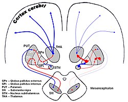
The image shows dopaminergic pathways of the human brain in normal condition (left) and Parkinsons Disease (right). Red Arrows indicate suppression of the target, blue arrows indicate stimulation of target structure. (Thalamic fasciculus visible but not labeled, as red line from GPi to THA.)
Details
Identifiers
Latin fasciculus thalamicus
NeuroNames 439
TA A14.1.08.679
A14.1.09.523
FMA 62065
Anatomical terms of neuroanatomy

The thalamic fasciculus is a component of the subthalamus. It is synonymous with field H1 of Forel. Nerve fibres form a tract containing cerebellothalamic (crossed) and pallidothalamic (uncrossed) fibres, that is insinuated between the thalamus and the zona incerta.

Subthalamus part of diencephalon

The subthalamus or prethalamus is a part of the diencephalon. Its most prominent structure is the subthalamic nucleus. The subthalamus connects to the globus pallidus, a basal nucleus of the telencephalon.

The fields of Forel are areas in a deep part of the brain known as the diencephalon. They are below the thalamus and consist of three defined, white matter areas of the subthalamus. These three regions are also named "H fields":

Axon The long process of a neuron that conducts nerve impulses, usually away from the cell body to the terminals and varicosities, which are sites of storage and release of neurotransmitter.

An axon, or nerve fiber, is a long, slender projection of a nerve cell, or neuron, in vertebrates, that typically conducts electrical impulses known as action potentials away from the nerve cell body. The function of the axon is to transmit information to different neurons, muscles, and glands. In certain sensory neurons, such as those for touch and warmth, the axons are called afferent nerve fibers and the electrical impulse travels along these from the periphery to the cell body, and from the cell body to the spinal cord along another branch of the same axon. Axon dysfunction has caused many inherited and acquired neurological disorders which can affect both the peripheral and central neurons. Nerve fibers are classed into three types – group A nerve fibers, group B nerve fibers, and group C nerve fibers. Groups A and B are myelinated, and group C are unmyelinated. These groups include both sensory fibers and motor fibers. Another classification groups only the sensory fibers as Type I, Type II, Type III, and Type IV.

The thalamic fasciculus consists of fibers from the ansa lenticularis and from the lenticular fasciculus, coming from different portions of the medial globus pallidus, before they jointly enter the ventral anterior nucleus of the thalamus. [1]

Ansa lenticularis

The ansa lenticularis is a part of the brain, making up the superior layer of the substantia innominata. Its fibers, derived from the medullary lamina of the lentiform nucleus, pass medially to end in the thalamus and subthalamic region, while others are said to end in the tegmentum and red nucleus. It is classified by NeuroNames as part of the subthalamus.

Lenticular fasciculus

The lenticular fasciculus is a tract connecting the globus pallidus to the thalamic fasciculus. It is synonymous with field H2 of Forel. The thalamic fasciculus is (composed of the lenticular fasciculus and ansa lenticularis) runs into the thalamus. It connects the globus pallidus to the thalamus.

Ventral anterior nucleus

The ventral anterior nucleus (VA) is a nucleus of the thalamus. It acts with the anterior part of the ventral lateral nucleus to modify signals from the basal ganglia.

Related Research Articles

Thalamus part of diencephalon, which is in turn part of prosencephalon (forebrain)

The thalamus is a large mass of gray matter in the dorsal part of the diencephalon of the brain with several functions such as relaying of sensory signals, including motor signals to the cerebral cortex, and the regulation of consciousness, sleep, and alertness.

Mammillary body pair of small round bodies, located on the undersurface of the brain that, as part of the diencephalon, form part of the limbic system

The mammillary bodies are a pair of small round bodies, located on the undersurface of the brain that, as part of the diencephalon, form part of the limbic system. They are located at the ends of the anterior arches of the fornix. They consist of two groups of nuclei, the medial mammillary nuclei and the lateral mammillary nuclei.

In the anatomy of the brain, the centromedian nucleus, also known as the centrum medianum, is a part of the intralaminar nucleus (ILN) of the thalamus. There are two centromedian nuclei arranged bilaterally.

Epithalamus part of diencephalon

The epithalamus is a (dorsal) posterior segment of the diencephalon. The diencephalon is a part of the forebrain that also contains the thalamus, the hypothalamus and pituitary gland. The epithalamus includes the habenula and their interconnecting fibers, the habenular commissure, the stria medullaris and the pineal gland.

Vestibular nuclei cranial nuclei for the vestibular nerve

The vestibular nuclei (VN) are the cranial nuclei for the vestibular nerve.

The mammillothalamic tract arises from cells in both the medial and lateral nuclei of the mammillary body and by fibers that are directly continued from the fornix.

Superior cerebellar peduncle

In the human brain, the superior cerebellar peduncle is a paired structure of white matter that connects the cerebellum to the midbrain. It consists mainly of efferent fibers, the cerebellothalamic tract that runs from a cerebellar hemisphere to the contralateral thalamus, and the cerebellorubral tract that runs from a cerebellar hemisphere to the red nucleus. It also contains afferent tracts, most prominent of which is the ventral spinocerebellar tract. Other afferent tracts are the trigeminothalamic fibers, tectocerebellar fibers, and noradrenergic fibers from the locus coeruleus. The superior peduncle emerges from the upper and medial parts of the white matter of each hemisphere and is placed under cover of the upper part of the cerebellum.

Ventral lateral nucleus

The ventral lateral nucleus (VL) is a nucleus in the ventral nuclear group of the thalamus.

Emboliform nucleus

The emboliform nucleus is a deep cerebellar nucleus that lies immediately to the medial side of the nucleus dentatus, and partly covering its hilum. It is one among the four pairs of deep cerebellar nuclei, which are from lateral to medial: the dentate, interposed, and fastigial nuclei. These nuclei can be seen using Weigert's elastic stain.

Basilar part of pons

The basilar part of pons is the ventral part of the pons; the dorsal part is known as the pontine tegmentum.

Rostral interstitial nucleus of medial longitudinal fasciculus

The rostral interstitial nucleus of medial longitudinal fasciculus (riMLF) is a portion of the medial longitudinal fasciculus which controls vertical gaze.

Posterior cerebral artery syndrome Human disease

Posterior cerebral artery syndrome is a condition whereby the blood supply from the posterior cerebral artery (PCA) is restricted, leading to a reduction of the function of the portions of the brain supplied by that vessel: the occipital lobe, the inferomedial temporal lobe, a large portion of the thalamus, and the upper brainstem and midbrain.

The dentatothalamic tract is a tract which connects the dentate nucleus and the thalamus while sending collaterals to the red nucleus.

Subthalamic fasciculus

The subthalamic fasciculus is a tract which connects the subthalamic nucleus (STN) and the globus pallidus (GP).

Central tegmental tract

The central tegmental tract is a structure in the midbrain and pons.

Pallidothalamic tracts

The pallidothalamic tracts are a part of the basal ganglia. They provide connectivity between the internal globus pallidus (GPi) and the thalamus, primarily the ventral anterior nucleus and the ventral lateral nucleus.

References

  1. Estomih Mtui; Gregory Gruener (2006). Clinical Neuroanatomy and Neuroscience: With STUDENT CONSULT Online Access. Philadelphia: Saunders. p. 359. ISBN   1-4160-3445-5.