Medial geniculate nucleus

Last updated
Medial geniculate nucleus
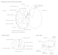
ThalamicNuclei.svg
Aud pathway.png
Auditory pathway (Medial geniculate body labeled at upper right, second from top)
Details
Part of Thalamus
System Auditory system
Artery Striate
Identifiers
Latin corpus geniculatum mediale
NeuroNames 355
NeuroLex ID birnlex_1670
TA98 A14.1.08.303
A14.1.08.808
TA2 5667, 5702
FMA 62211
Anatomical terms of neuroanatomy

The medial geniculate nucleus (MGN) or medial geniculate body (MGB) is part of the auditory thalamus and represents the thalamic relay between the inferior colliculus (IC) and the auditory cortex (AC). It is made up of a number of sub-nuclei that are distinguished by their neuronal morphology and density, by their afferent and efferent connections, and by the coding properties of their neurons. It is thought that the MGN influences the direction and maintenance of attention.
Contents

Divisions

The MGN has three major divisions; ventral (VMGN), dorsal (DMGN) and medial (MMGN). Whilst the VMGN is specific to auditory information processing, the DMGN and MMGN also receive information from non-auditory pathways.

DivisionInputsOutputs
VMGN* Inferior colliculus
** ICC (ipsilateral)
** ICP (contralateral)
* Reticular nucleus of the thalamus (ipsilateral)
* Auditory cortex
Auditory cortex
* Anterior (AAF)
* Primary (AI)
* Posterior (PAF)
DMGN* IC
** Pericentral nuclei
** External nuclei
* Auditory cortex
* Other thalamic nuclei
Auditory cortex
MMGN* ICC (ipsilateral)
* LLN (ipsi and contra)
* Superior colliculus
* Periolivary nuclei
* Auditory cortex
** Secondary (AII)
* Reticular nucleus of thalamus
* Somatosensory and vestibular influences are also present
Auditory Cortex
* AII (ipsilateral)
* AAF (ipsilateral)
* AI (ipsilateral)
* PAF (ipsilateral)

Ventral subnucleus

Cell types

There are two main cell types in the ventral subnucleus of the medial geniculate body (VMGN):

  • Thalamocortical relay cells (or principal neurons): The dendritic input to these cells comes from two sets of dendritic trees oriented on opposite poles of the cell. The long axis of the relay cells lie parallel to each other running superior-inferiorly with the dendritic trees of cells within the same iso-frequency band overlapping. This is similar to the dendritic organization of the IC, but with a different orientation. The dendrites of relay cells form a synaptic nest with ascending axons from the inferior colliculus and intrathalamic interneurons. In this synapse, relay cells are excited by input from the IC axons. At the same time, they are inhibited by dendritic synapses from the interneurons of the VMGB. This type of synaptic nesting is characteristic of other regions in the thalamus as well.
  • Intrathalamic Interneurons: These interneurons provide inhibitory (GABA) input to the relay cells at the synaptic nests. The target of their axons however, is not clear. Some interneurons appear to target relay cells, while others target other interneurons. There is also at least one type of interneuron that appears to not be involved in the synaptic nests.

Function

The VMGN is thought to be primarily responsible for relaying frequency, intensity and binaural information to the cortex. The responses in the VMGN appear to be organized in a tonotopically similar way to those in the IC. The primary difference being that the iso-frequency bands are arranged such that lateral regions are most responsive to low frequencies and medial regions are responsive to high frequencies. Spatiotopic and modulotopic maps (as in the IC) however have not been well supported by mammalian studies. Both monaural (10%) and binaural cells (90%) exist in the MGN. The monaural cells are primarily responsive to sound in the contralateral hemifield. Binaural cells are typically similar to the EE or EI type found in the IC.

Definitions of abbreviations

IC = Inferior colliculus

EE (Excitatory excitatory) type neurons are characterized by excitatory responses to monaural stimulations of both ears. This response may either be higher than the monaural response (EE– facilitation) Or lower (EE– occlusion)

EI (Excitatory inhibitory) type neurons Are characterized by monaural excitation (usually from the contralateral ear). The ipsilateral neuron is inhibited when contralateral ear is stimulated at the same time

Dorsal subnucleus

Cell types

There are a large number of cell types present in the dorsal subnucleus of the medial geniculate body (DMGN):

At least two principal cell types have been found, along with two distinct types of interneurons. Several sub-nuclei have been identified based on morphology. No frequency-specific layering has been found in the DMGN.

Function

Many types of responses are present in the DMGB that appear to vary by sub-nuclei. Generally, the responses are broadly tuned, but some cells appear to respond only to complex stimuli. Other cells are multi modal, often responding to somatosensory as well as auditory stimuli.

Medial subnucleus

Cell types

Cells in the medial subnucleus of the medial geniculate body (MMGN) have large irregular shaped dendritic trees. There is no clear segregation based on the source of these inputs.

Function

The MMGN seems to functionally be responsible for detection of the relative intensity and duration of a sound. It shows a wide range of responses to auditory stimuli. Binaural interactions found in the MMGN include EE, EI, and IE types. Both broadly and narrowly tuned cells have been observed. A type of intensity tuning has also been observed. In this type of cell, the response actually decreases as sound intensity increases above a specific level. Almost all cells in the MMGN appear to respond for the duration of the stimulus, and have very little adaptation. Individual cells still appear to be preferentially tuned to certain frequencies, but they often have more than one and are broadly tuned within these cell frequencies. It is not clear whether there truly is one, none, or many tonotopic organizations maps present in the MMGN. Anaesthetics tend to have large effects on cells within the MMGN, making responses difficult to study. Finally, the behaviour of MMGN cells are complicated by the fact that sensory stimulation from other modalities modifies the responsiveness of many, but not all, cells in the MMGN.

Additional images

Related Research Articles

Articles related to anatomy include:

<span class="mw-page-title-main">Lateral geniculate nucleus</span> Component of the visual system in the brains thalamus

In neuroanatomy, the lateral geniculate nucleus is a structure in the thalamus and a key component of the mammalian visual pathway. It is a small, ovoid, ventral projection of the thalamus where the thalamus connects with the optic nerve. There are two LGNs, one on the left and another on the right side of the thalamus. In humans, both LGNs have six layers of neurons alternating with optic fibers.

<span class="mw-page-title-main">Auditory system</span> Sensory system used for hearing

The auditory system is the sensory system for the sense of hearing. It includes both the sensory organs and the auditory parts of the sensory system.

<span class="mw-page-title-main">Lateral lemniscus</span>

The lateral lemniscus is a tract of axons in the brainstem that carries information about sound from the cochlear nucleus to various brainstem nuclei and ultimately the contralateral inferior colliculus of the midbrain. Three distinct, primarily inhibitory, cellular groups are located interspersed within these fibers, and are thus named the nuclei of the lateral lemniscus.

<span class="mw-page-title-main">Inferior colliculus</span> Midbrain structure involved in the auditory pathway

The inferior colliculus (IC) is the principal midbrain nucleus of the auditory pathway and receives input from several peripheral brainstem nuclei in the auditory pathway, as well as inputs from the auditory cortex. The inferior colliculus has three subdivisions: the central nucleus, a dorsal cortex by which it is surrounded, and an external cortex which is located laterally. Its bimodal neurons are implicated in auditory-somatosensory interaction, receiving projections from somatosensory nuclei. This multisensory integration may underlie a filtering of self-effected sounds from vocalization, chewing, or respiration activities.

<span class="mw-page-title-main">Koniocellular cell</span>

A koniocellular cell is a neuron with a small cell body that is located in the koniocellular layer of the lateral geniculate nucleus (LGN) in primates, including humans.

<span class="mw-page-title-main">Thalamocortical radiations</span> Neural pathways between the thalamus and cerebral cortex

In neuroanatomy, thalamocortical radiations, also known as thalamocortical fibres, are the efferent fibres that project from the thalamus to distinct areas of the cerebral cortex. They form fibre bundles that emerge from the lateral surface of the thalamus.

<span class="mw-page-title-main">Golgi cell</span>

In neuroscience, Golgi cells are the most abundant inhibitory interneurons found within the granular layer of the cerebellum. Golgi cells can be found in the granular layer at various layers. The Golgi cell is essential for controlling the activity of the granular layer. They were first identified as inhibitory in 1964. It was also the first example of an inhibitory feedback network in which the inhibitory interneuron was identified anatomically. Golgi cells produce a wide lateral inhibition that reaches beyond the afferent synaptic field and inhibit granule cells via feedforward and feedback inhibitory loops. These cells synapse onto the dendrite of granule cells and unipolar brush cells. They receive excitatory input from mossy fibres, also synapsing on granule cells, and parallel fibers, which are long granule cell axons. Thereby this circuitry allows for feed-forward and feed-back inhibition of granule cells.

<span class="mw-page-title-main">Cochlear nucleus</span> Two cranial nerve nuclei of the human brainstem

The cochlear nuclear (CN) complex comprises two cranial nerve nuclei in the human brainstem, the ventral cochlear nucleus (VCN) and the dorsal cochlear nucleus (DCN). The ventral cochlear nucleus is unlayered whereas the dorsal cochlear nucleus is layered. Auditory nerve fibers, fibers that travel through the auditory nerve carry information from the inner ear, the cochlea, on the same side of the head, to the nerve root in the ventral cochlear nucleus. At the nerve root the fibers branch to innervate the ventral cochlear nucleus and the deep layer of the dorsal cochlear nucleus. All acoustic information thus enters the brain through the cochlear nuclei, where the processing of acoustic information begins. The outputs from the cochlear nuclei are received in higher regions of the auditory brainstem.

<span class="mw-page-title-main">Dorsal cochlear nucleus</span>

The dorsal cochlear nucleus is a cortex-like structure on the dorso-lateral surface of the brainstem. Along with the ventral cochlear nucleus (VCN), it forms the cochlear nucleus (CN), where all auditory nerve fibers from the cochlea form their first synapses.

<span class="mw-page-title-main">Superior olivary complex</span> Collection of brainstem nuclei related to hearing

The superior olivary complex (SOC) or superior olive is a collection of brainstem nuclei that is located in pons, functions in multiple aspects of hearing and is an important component of the ascending and descending auditory pathways of the auditory system. The SOC is intimately related to the trapezoid body: most of the cell groups of the SOC are dorsal to this axon bundle while a number of cell groups are embedded in the trapezoid body. Overall, the SOC displays a significant interspecies variation, being largest in bats and rodents and smaller in primates.

<span class="mw-page-title-main">Interaural time difference</span> Difference in time that it takes a sound to travel between two ears

The interaural time difference when concerning humans or animals, is the difference in arrival time of a sound between two ears. It is important in the localization of sounds, as it provides a cue to the direction or angle of the sound source from the head. If a signal arrives at the head from one side, the signal has further to travel to reach the far ear than the near ear. This pathlength difference results in a time difference between the sound's arrivals at the ears, which is detected and aids the process of identifying the direction of sound source.

A topographic map is the ordered projection of a sensory surface, like the retina or the skin, or an effector system, like the musculature, to one or more structures of the central nervous system. Topographic maps can be found in all sensory systems and in many motor systems.

Binaural fusion or binaural integration is a cognitive process that involves the combination of different auditory information presented binaurally, or to each ear. In humans, this process is essential in understanding speech as one ear may pick up more information about the speech stimuli than the other.

The isothalamus is a division used by some researchers in describing the thalamus.

<span class="mw-page-title-main">Synaptic gating</span>

Synaptic gating is the ability of neural circuits to gate inputs by either suppressing or facilitating specific synaptic activity. Selective inhibition of certain synapses has been studied thoroughly, and recent studies have supported the existence of permissively gated synaptic transmission. In general, synaptic gating involves a mechanism of central control over neuronal output. It includes a sort of gatekeeper neuron, which has the ability to influence transmission of information to selected targets independently of the parts of the synapse upon which it exerts its action.

<span class="mw-page-title-main">Ventral cochlear nucleus</span>

In the ventral cochlear nucleus (VCN), auditory nerve fibers enter the brain via the nerve root in the VCN. The ventral cochlear nucleus is divided into the anterior ventral (anteroventral) cochlear nucleus (AVCN) and the posterior ventral (posteroventral) cochlear nucleus (PVCN). In the VCN, auditory nerve fibers bifurcate, the ascending branch innervates the AVCN and the descending branch innervates the PVCN and then continue to the dorsal cochlear nucleus. The orderly innervation by auditory nerve fibers gives the AVCN a tonotopic organization along the dorsoventral axis. Fibers that carry information from the apex of the cochlea that are tuned to low frequencies contact neurons in the ventral part of the AVCN; those that carry information from the base of the cochlea that are tuned to high frequencies contact neurons in the dorsal part of the AVCN. Several populations of neurons populate the AVCN. Bushy cells receive input from auditory nerve fibers through particularly large endings called end bulbs of Held. They contact stellate cells through more conventional boutons.

Low-threshold spikes (LTS) refer to membrane depolarizations by the T-type calcium channel. LTS occur at low, negative, membrane depolarizations. They often follow a membrane hyperpolarization, which can be the result of decreased excitability or increased inhibition. LTS result in the neuron reaching the threshold for an action potential. LTS is a large depolarization due to an increase in Ca2+ conductance, so LTS is mediated by calcium (Ca2+) conductance. The spike is typically crowned by a burst of two to seven action potentials, which is known as a low-threshold burst. LTS are voltage dependent and are inactivated if the cell's resting membrane potential is more depolarized than −60mV. LTS are deinactivated, or recover from inactivation, if the cell is hyperpolarized and can be activated by depolarizing inputs, such as excitatory postsynaptic potentials (EPSP). LTS were discovered by Rodolfo Llinás and coworkers in the 1980s.

The olivocochlear system is a component of the auditory system involved with the descending control of the cochlea. Its nerve fibres, the olivocochlear bundle (OCB), form part of the vestibulocochlear nerve, and project from the superior olivary complex in the brainstem (pons) to the cochlea.

The Mauthner cells are a pair of big and easily identifiable neurons located in the rhombomere 4 of the hindbrain in fish and amphibians that are responsible for a very fast escape reflex. The cells are also notable for their unusual use of both chemical and electrical synapses.

References