Vestibulocochlear nerve

Last updated
Vestibulocochlear nerve
Gray789.png
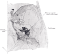
The course and connections of the facial nerve in the temporal bone
Brain human normal inferior view with labels en.svg
Inferior view of the human brain, with the cranial nerves labelled.
Details
To Cochlear nerve, vestibular nerve
Innervates Hearing, balance
Identifiers
Latin nervus vestibulocochlearis
MeSH D000159
NeuroNames 553
TA98 A14.2.01.121
TA2 6307
FMA 50869
Anatomical terms of neuroanatomy

The vestibulocochlear nerve or auditory vestibular nerve, also known as the eighth cranial nerve, cranial nerve VIII, or simply CN VIII, is a cranial nerve that transmits sound and equilibrium (balance) information from the inner ear to the brain. Through olivocochlear fibers, it also transmits motor and modulatory information from the superior olivary complex in the brainstem to the cochlea. [1]

Contents

Structure

The vestibulocochlear nerve consists mostly of bipolar neurons and splits into two large divisions: the cochlear nerve and the vestibular nerve.

Cranial nerve 8, the vestibulocochlear nerve, goes to the middle portion of the brainstem called the pons (which then is largely composed of fibers going to the cerebellum). The 8th cranial nerve runs between the base of the pons and medulla oblongata (the lower portion of the brainstem). This junction between the pons, medulla, and cerebellum that contains the 8th nerve is called the cerebellopontine angle. The vestibulocochlear nerve is accompanied by the labyrinthine artery, which usually branches off from the anterior inferior cerebellar artery at the cerebellopontine angle, and then goes with the 7th nerve through the internal acoustic meatus to the internal ear.

The cochlear nerve travels away from the cochlea of the inner ear where it starts as the spiral ganglia. Processes from the organ of Corti conduct afferent transmission to the spiral ganglia. It is the inner hair cells of the organ of Corti that are responsible for activation of afferent receptors in response to pressure waves reaching the basilar membrane through the transduction of sound. The exact mechanism by which sound is transmitted by the neurons of the cochlear nerve is uncertain; the two competing theories are place theory and temporal theory.

The vestibular nerve travels from the vestibular system of the inner ear. The vestibular ganglion houses the cell bodies of the bipolar neurons and extends processes to five sensory organs. Three of these are the cristae located in the ampullae of the semicircular canals. Hair cells of the cristae activate afferent receptors in response to rotational acceleration. The other two sensory organs supplied by the vestibular neurons are the maculae of the saccule and utricle. Hair cells of the maculae in the utricle activate afferent receptors in response to linear acceleration, while hair cells of the maculae in the saccule respond to vertically directed linear force.

Development

The vestibulocochlear nerve is derived from the embryonic otic placode.

Function

This is the nerve along which the sensory cells (the hair cells) of the inner ear transmit information to the brain. It consists of the cochlear nerve, carrying details about hearing, and the vestibular nerve, carrying information about balance. It emerges from the pontomedullary junction and exits the inner skull via the internal acoustic meatus in the temporal bone.

The vestibulocochlear nerve carries axons of type special somatic afferent.

Clinical significance

Symptoms of damage

Damage to the vestibulocochlear nerve may cause the following symptoms:

Examination

Examinations that can be done include the Rinne and Weber tests.

Rinne's test involves Rinne's Right and Left Test, since auditory acuity is equal in both ears. If bone conduction (BC) is more than air conduction (AC) (BC>AC) indicates Rinne Test is negative or abnormal. If AC>BC Rinne test is normal or positive. If BC>AC and Weber's test lateralizes to abnormal side then it is Conductive hearing loss. If AC>BC and Weber's test lateralizes to normal side then it concludes Sensorineural hearing loss.

After pure-tone testing, if the AC and BC responses at all frequencies 500–8000 Hz are better than 25 dB HL, meaning 0-24 dB HL, the results are considered normal hearing sensitivity. If the AC and BC are worse than 25 dB HL at any one or more frequency between 500 and 8000 Hz, meaning 25+, and there is a no bigger difference between AC and BC beyond 10 dB at any frequency, there is a sensori-neural hearing loss present. If the BC responses are normal, 0-24 dB HL, and the AC are worse than 25 dB HL, as well as a 10 dB gap between the air and bone responses, a conductive hearing loss is present. {updated March 2019}

The modified Hughson–Westlake method is used by many audiologists during testing. A battery of (1) otoscopy, to view the ear canal and tympanic membrane, (2) tympanometry, to assess the immittance of the tympanic membrane and how well it moves, (3) otoacoustic emissions, to measure the response of the outer hair cells located in the cochlea, (4) audiobooth pure-tone testing, to obtain thresholds to determine the type, severity, and pathology of the hearing loss present, and (5) speech tests, to measure the patients recognition and ability to repeat the speech heard, is all taken into consideration when diagnosing the pathology of the patient.

History

Etymology

Some older texts call the nerve the acoustic or auditory nerve, [3] but these terms have fallen out of widespread use because they fail to recognize the nerve's role in the vestibular system. Vestibulocochlear nerve is therefore preferred by most.

See also

Related Research Articles

<span class="mw-page-title-main">Inner ear</span> Innermost part of the vertebrate ear

The inner ear is the innermost part of the vertebrate ear. In vertebrates, the inner ear is mainly responsible for sound detection and balance. In mammals, it consists of the bony labyrinth, a hollow cavity in the temporal bone of the skull with a system of passages comprising two main functional parts:

<span class="mw-page-title-main">Cochlea</span> Snail-shaped part of inner ear involved in hearing

The cochlea is the part of the inner ear involved in hearing. It is a spiral-shaped cavity in the bony labyrinth, in humans making 2.75 turns around its axis, the modiolus. A core component of the cochlea is the organ of Corti, the sensory organ of hearing, which is distributed along the partition separating the fluid chambers in the coiled tapered tube of the cochlea.

<span class="mw-page-title-main">Pons</span> Part of the brainstem in humans and other bipeds

The pons is part of the brainstem that in humans and mammals, lies inferior to the midbrain, superior to the medulla oblongata and anterior to the cerebellum.

<span class="mw-page-title-main">Brainstem</span> Posterior part of the brain, adjoining and structurally continuous

The brainstem is the stalk-like part of the brain that interconnects the cerebrum and diencephalon with the spinal cord. In the human brain, the brainstem is composed of the midbrain, the pons, and the medulla oblongata. The midbrain is continuous with the thalamus of the diencephalon through the tentorial notch.

<span class="mw-page-title-main">Saccule</span> Bed of sensory cells in the inner ear

The saccule is a bed of sensory cells in the inner ear. It translates head movements into neural impulses for the brain to interpret. The saccule detects linear accelerations and head tilts in the vertical plane. When the head moves vertically, the sensory cells of the saccule are disturbed and the neurons connected to them begin transmitting impulses to the brain. These impulses travel along the vestibular portion of the eighth cranial nerve to the vestibular nuclei in the brainstem.

<span class="mw-page-title-main">Organ of Corti</span> Receptor organ for hearing

The organ of Corti, or spiral organ, is the receptor organ for hearing and is located in the mammalian cochlea. This highly varied strip of epithelial cells allows for transduction of auditory signals into nerve impulses' action potential. Transduction occurs through vibrations of structures in the inner ear causing displacement of cochlear fluid and movement of hair cells at the organ of Corti to produce electrochemical signals.

<span class="mw-page-title-main">Auditory system</span> Sensory system used for hearing

The auditory system is the sensory system for the sense of hearing. It includes both the sensory organs and the auditory parts of the sensory system.

<span class="mw-page-title-main">Hair cell</span> Auditory sensory receptor nerve cells

Hair cells are the sensory receptors of both the auditory system and the vestibular system in the ears of all vertebrates, and in the lateral line organ of fishes. Through mechanotransduction, hair cells detect movement in their environment.

<span class="mw-page-title-main">Sensorineural hearing loss</span> Hearing loss caused by an inner ear or vestibulocochlear nerve defect

Sensorineural hearing loss (SNHL) is a type of hearing loss in which the root cause lies in the inner ear or sensory organ or the vestibulocochlear nerve. SNHL accounts for about 90% of reported hearing loss. SNHL is usually permanent and can be mild, moderate, severe, profound, or total. Various other descriptors can be used depending on the shape of the audiogram, such as high frequency, low frequency, U-shaped, notched, peaked, or flat.

Auditory neuropathy (AN) is a hearing disorder in which the outer hair cells of the cochlea are present and functional, but sound information is not transmitted sufficiently by the auditory nerve to the brain. Hearing loss with AN can range from normal hearing sensitivity to profound hearing loss.

<span class="mw-page-title-main">Stereocilia (inner ear)</span> Mechanosensing organelles of hair cells

In the inner ear, stereocilia are the mechanosensing organelles of hair cells, which respond to fluid motion in numerous types of animals for various functions, including hearing and balance. They are about 10–50 micrometers in length and share some similar features of microvilli. The hair cells turn the fluid pressure and other mechanical stimuli into electric stimuli via the many microvilli that make up stereocilia rods. Stereocilia exist in the auditory and vestibular systems.

<span class="mw-page-title-main">Cochlear nerve</span> Nerve carrying auditory information from the inner ear to the brain

The cochlear nerve is one of two parts of the vestibulocochlear nerve, a cranial nerve present in amniotes, the other part being the vestibular nerve. The cochlear nerve carries auditory sensory information from the cochlea of the inner ear directly to the brain. The other portion of the vestibulocochlear nerve is the vestibular nerve, which carries spatial orientation information to the brain from the semicircular canals, also known as semicircular ducts.

<span class="mw-page-title-main">Cochlear nucleus</span> Two cranial nerve nuclei of the human brainstem

The cochlear nuclear (CN) complex comprises two cranial nerve nuclei in the human brainstem, the ventral cochlear nucleus (VCN) and the dorsal cochlear nucleus (DCN). The ventral cochlear nucleus is unlayered whereas the dorsal cochlear nucleus is layered. Auditory nerve fibers, fibers that travel through the auditory nerve carry information from the inner ear, the cochlea, on the same side of the head, to the nerve root in the ventral cochlear nucleus. At the nerve root the fibers branch to innervate the ventral cochlear nucleus and the deep layer of the dorsal cochlear nucleus. All acoustic information thus enters the brain through the cochlear nuclei, where the processing of acoustic information begins. The outputs from the cochlear nuclei are received in higher regions of the auditory brainstem.

The vestibular ganglion is a collection of cell bodies belonging to first order sensory neurons of the vestibular nerve. It is located within the internal auditory canal.

<span class="mw-page-title-main">Spiral ganglion</span> Anatomical structure

The spiral (cochlear) ganglion is a group of neuron cell bodies in the modiolus, the conical central axis of the cochlea. These bipolar neurons innervate the hair cells of the organ of Corti. They project their axons to the ventral and dorsal cochlear nuclei as the cochlear nerve, a branch of the vestibulocochlear nerve.

Binaural fusion or binaural integration is a cognitive process that involves the combination of different auditory information presented binaurally, or to each ear. In humans, this process is essential in understanding speech as one ear may pick up more information about the speech stimuli than the other.

The cerebellopontine angle syndrome is a distinct neurological syndrome of deficits that can arise due to the closeness of the cerebellopontine angle to specific cranial nerves. Indications include unilateral hearing loss (85%), speech impediments, disequilibrium, tremors or other loss of motor control. The cerebellopontine angle cistern is a subarachnoid cistern formed by the cerebellopontine angle that lies between the cerebellum and the pons. It is filled with cerebrospinal fluid and is a common site for the growth of acoustic neuromas or schwannomas.

The olivocochlear system is a component of the auditory system involved with the descending control of the cochlea. Its nerve fibres, the olivocochlear bundle (OCB), form part of the vestibulocochlear nerve, and project from the superior olivary complex in the brainstem (pons) to the cochlea.

The neural encoding of sound is the representation of auditory sensation and perception in the nervous system. The complexities of contemporary neuroscience are continually redefined. Thus what is known of the auditory system has been continually changing. The encoding of sounds includes the transduction of sound waves into electrical impulses along auditory nerve fibers, and further processing in the brain.

Electrocochleography is a technique of recording electrical potentials generated in the inner ear and auditory nerve in response to sound stimulation, using an electrode placed in the ear canal or tympanic membrane. The test is performed by an otologist or audiologist with specialized training, and is used for detection of elevated inner ear pressure or for the testing and monitoring of inner ear and auditory nerve function during surgery.

References

  1. Lopez-Poveda, Enrique A. (26 March 2018). "Olivocochlear Efferents in Animals and Humans: From Anatomy to Clinical Relevance". Frontiers in Neurology. 9: 197. doi: 10.3389/fneur.2018.00197 . PMC   5879449 . PMID   29632514.
  2. Coad, ML; Lockwood, A; Salvi, R; Burkard, R (2001). "Characteristics of patients with gaze-evoked tinnitus". Otology & Neurotology. 22 (5): 650–4. doi:10.1097/00129492-200109000-00016. PMID   11568674. S2CID   44391826.
  3. "IX. Neurology. 5h. The Acoustic Nerve. Gray, Henry. 1918. Anatomy of the Human Body". www.bartleby.com.

Additional images