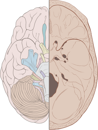
Terminal nerve

Last updated
Terminal nerve
Lawrence 1960 1.3.png
Left The terminal nerve as it is shown on the ventral side of a dog-fish brain. (Topmost label)
Details
Identifiers
Latin nervus terminalis
TA98 A14.2.01.002
TA2 6179
FMA 76749
Anatomical terminology

The terminal nerve, also known as cranial nerve zero or simply as CN 0, is a nerve that was not included in the seminal classification of the cranial nerves as CN I through CN XII, but has since been recognized and listed in TA2. [1] It was discovered by German scientist Gustav Fritsch in 1878 in the brains of sharks, and was first found in humans in 1913. [2] Studies have confirmed that the terminal nerve is a common finding in the adult human brain. [3] [4]

Contents

The accepted name of terminal nerve is due to its entrance in the lamina terminalis regions. [1] The nerve has previously been called cranial nerve XIII, zero nerve, nerve N, and NT. [5] [6]

Structure

The original images (1878) of Fritsch's dogfish shark brain showing the nerve marked by an asterisk Fritsch dogfish shark brain.png
The original images (1878) of Fritsch's dogfish shark brain showing the nerve marked by an asterisk

The terminal nerve appears just in front of the other cranial nerves and would, if earlier recognized, have been classified as cranial nerve one. It first appears bilaterally as a microscopic plexus of unmyelinated peripheral nerve fibers in the subarachnoid space covering the straight gyrus. The plexus appears near the cribriform plate and travels posteriorly toward the olfactory trigone and lamina terminalis. [3]

The terminal nerve is clearly seen in the human embryo but loses some of its ganglion cells before birth making it less recognizable in adults. [7] The nerve is therefore often overlooked in autopsies, and is often torn out upon exposing the brain. [5] Careful dissection is necessary to visualize the nerve.

Development

The zebrafish was used as a developmental model in research from 2004. [8]

The connections between the terminal nerve and the olfactory system have been extensively studied in human embryos. olfactory nerve fibers enter the brain at stage 17, fibers from the vomeronasal organ and fibers of the terminal nerve enter the brain at stages 17 and 18. [9] During prenatal development some of the ganglion cells are lost. [7]

Function

The functions of the terminal nerve are only speculated on together with possible pathological implications. [1] Although very close to the olfactory nerve, the terminal nerve is not connected to the olfactory bulb, where smells are analyzed. This suggests that the nerve is either vestigial or may be related to the sensing of pheromones. [10] The nerve may modulate olfactory inputs making pheromones (particularly sex pheromones) more detectable. [1] This hypothesis is further supported by the fact that the terminal nerve projects to the medial and lateral septal nuclei and the preoptic areas, all of which are involved in reproduction in mammals. [2] A 1987 study found that mating in hamsters is reduced when the terminal nerve is severed. [11]

Clinical significance

Alterations in the terminal nerve structure may be implicated in Kallmann syndrome. [7] [12]

Additional images

Related Research Articles

<span class="mw-page-title-main">Cranial nerves</span> Nerves that emerge directly from the brain

Cranial nerves are the nerves that emerge directly from the brain, of which there are conventionally considered twelve pairs. Cranial nerves relay information between the brain and parts of the body, primarily to and from regions of the head and neck, including the special senses of vision, taste, smell, and hearing.

<span class="mw-page-title-main">Vagus nerve</span> Main nerve of the parasympathetic nervous system

The vagus nerve, also known as the tenth cranial nerve, cranial nerve X, or simply CN X, is a cranial nerve that carries sensory fibers that create a pathway that interfaces with the parasympathetic control of the heart, lungs, and digestive tract.

<span class="mw-page-title-main">Optic nerve</span> Second cranial nerve, which connects the eyes to the brain

In neuroanatomy, the optic nerve, also known as the second cranial nerve, cranial nerve II, or simply CN II, is a paired cranial nerve that transmits visual information from the retina to the brain. In humans, the optic nerve is derived from optic stalks during the seventh week of development and is composed of retinal ganglion cell axons and glial cells; it extends from the optic disc to the optic chiasma and continues as the optic tract to the lateral geniculate nucleus, pretectal nuclei, and superior colliculus.

<span class="mw-page-title-main">Parasympathetic nervous system</span> Division of the autonomic nervous system

The parasympathetic nervous system (PSNS) is one of the three divisions of the autonomic nervous system, the others being the sympathetic nervous system and the enteric nervous system.

<span class="mw-page-title-main">Olfactory nerve</span> Cranial nerve I, for smelling

The olfactory nerve, also known as the first cranial nerve, cranial nerve I, or simply CN I, is a cranial nerve that contains sensory nerve fibers relating to the sense of smell.

<span class="mw-page-title-main">Facial nerve</span> Cranial nerve VII, for the face and tasting

The facial nerve, also known as the seventh cranial nerve, cranial nerve VII, or simply CN VII, is a cranial nerve that emerges from the pons of the brainstem, controls the muscles of facial expression, and functions in the conveyance of taste sensations from the anterior two-thirds of the tongue. The nerve typically travels from the pons through the facial canal in the temporal bone and exits the skull at the stylomastoid foramen. It arises from the brainstem from an area posterior to the cranial nerve VI and anterior to cranial nerve VIII.

Articles related to anatomy include:

<span class="mw-page-title-main">Oculomotor nerve</span> Cranial nerve III, for eye movements

The oculomotor nerve, also known as the third cranial nerve, cranial nerve III, or simply CN III, is a cranial nerve that enters the orbit through the superior orbital fissure and innervates extraocular muscles that enable most movements of the eye and that raise the eyelid. The nerve also contains fibers that innervate the intrinsic eye muscles that enable pupillary constriction and accommodation. The oculomotor nerve is derived from the basal plate of the embryonic midbrain. Cranial nerves IV and VI also participate in control of eye movement.

<span class="mw-page-title-main">Trigeminal nerve</span> Cranial nerve responsible for the faces senses and motor functions

In neuroanatomy, the trigeminal nerve (lit. triplet nerve), also known as the fifth cranial nerve, cranial nerve V, or simply CN V, is a cranial nerve responsible for sensation in the face and motor functions such as biting and chewing; it is the most complex of the cranial nerves. Its name (trigeminal, from Latin tri- 'three' and -geminus 'twin') derives from each of the two nerves (one on each side of the pons) having three major branches: the ophthalmic nerve (V1), the maxillary nerve (V2), and the mandibular nerve (V3). The ophthalmic and maxillary nerves are purely sensory, whereas the mandibular nerve supplies motor as well as sensory (or "cutaneous") functions. Adding to the complexity of this nerve is that autonomic nerve fibers as well as special sensory fibers (taste) are contained within it.

<span class="mw-page-title-main">Glossopharyngeal nerve</span> Cranial nerve IX, for the tongue and pharynx

The glossopharyngeal nerve, also known as the ninth cranial nerve, cranial nerve IX, or simply CN IX, is a cranial nerve that exits the brainstem from the sides of the upper medulla, just anterior to the vagus nerve. Being a mixed nerve (sensorimotor), it carries afferent sensory and efferent motor information. The motor division of the glossopharyngeal nerve is derived from the basal plate of the embryonic medulla oblongata, whereas the sensory division originates from the cranial neural crest.

<span class="mw-page-title-main">Vomeronasal organ</span> Smell sense organ above the roof of the mouth

The vomeronasal organ (VNO), or Jacobson's organ, is the paired auxiliary olfactory (smell) sense organ located in the soft tissue of the nasal septum, in the nasal cavity just above the roof of the mouth in various tetrapods. The name is derived from the fact that it lies adjacent to the unpaired vomer bone in the nasal septum. It is present and functional in all snakes and lizards, and in many mammals, including cats, dogs, cattle, pigs, and some primates. Some humans may have physical remnants of a VNO, but it is vestigial and non-functional.

<span class="mw-page-title-main">Mandibular nerve</span> Branch of the trigeminal nerve responsible for the lower face and jaw

In neuroanatomy, the mandibular nerve (V3) is the largest of the three divisions of the trigeminal nerve, the fifth cranial nerve (CN V). Unlike the other divisions of the trigeminal nerve (ophthalmic nerve, maxillary nerve) which contain only afferent fibers, the mandibular nerve contains both afferent and efferent fibers. These nerve fibers innervate structures of the lower jaw and face, such as the tongue, lower lip, and chin. The mandibular nerve also innervates the muscles of mastication.

<span class="mw-page-title-main">Olfactory system</span> Sensory system used for smelling

The olfactory system, or sense of smell, is the sensory system used for olfaction. Olfaction is one of the special senses directly associated with specific organs. Most mammals and reptiles have a main olfactory system and an accessory olfactory system. The main olfactory system detects airborne substances, while the accessory system senses fluid-phase stimuli.

<span class="mw-page-title-main">Edinger–Westphal nucleus</span> One of two nuclei of the oculomotor nerve

The Edinger–Westphal nucleus also called the accessory or visceral oculomotor nerve, is one of the two nuclei of the oculomotor nerve located in the midbrain. It receives afferents from both pretectal nuclei. It contains parasympathetic pre-ganglionic neuron cell bodies that synapse in the ciliary ganglion. It contributes the autonomic, parasympathetic component to the oculomotor nerve, ultimately providing innervation to the iris sphincter muscle and ciliary muscle to mediate the pupillary light reflex and accommodation, respectively.

<span class="mw-page-title-main">Lesser petrosal nerve</span> Nerve of the parotid gland

The lesser petrosal nerve is the general visceral efferent (GVE) nerve conveying pre-ganglionic parasympathetic secretomotor fibers for the parotid gland from the tympanic plexus to the otic ganglion. It passes out of the tympanic cavity through the petrous part of the temporal bone into the middle cranial fossa of the cranial cavity, then exits the cranial cavity through its own canaliculus to reach the infratemporal fossa.

<span class="mw-page-title-main">Olfactory tract</span> Part of the olfactory system

The olfactory tract is a bilateral bundle of afferent nerve fibers from the mitral and tufted cells of the olfactory bulb that connects to several target regions in the brain, including the piriform cortex, amygdala, and entorhinal cortex. It is a narrow white band, triangular on coronal section, the apex being directed upward.

The rostral neuropore or anterior neuropore is a region corresponding to the opening of the embryonic neural tube in the anterior portion of the developing prosencephalon. The central nervous system develops from the neural tube, which initially starts as a plate of cells in the ectoderm and this is called the neural plate, the neural plate then undergoes folding and starts closing from the center of the developing fetus, this leads to two open ends, one situated cranially/rostrally and the other caudally. Bending of the neural plate begins on day 22, and the cranial neuropore closes on day 24, giving rise to the lamina terminalis of the brain.

<span class="mw-page-title-main">Outline of the human nervous system</span> Overview of and topical guide to the human nervous system

The following diagram is provided as an overview of and topical guide to the human nervous system:

References

  1. 1 2 3 4 Ruqa WA, Pennacchia F, Rusi E, Zoccali F, Bruno G, Talarico G, Barbato C, Minni A (March 2024). "Smelling TNT: Trends of the Terminal Nerve". Int J Mol Sci. 25 (7): 3920. doi: 10.3390/ijms25073920 . PMC   11011448 . PMID   38612730.
  2. 1 2 Fields, R. Douglas (2007). "Sex and the Secret Nerve". Scientific American Mind. 18: 20–7. doi:10.1038/scientificamericanmind0207-20.
  3. 1 2 Fuller GN, Burger PC (1990). "Nervus terminalis (cranial nerve zero) in the adult human". Clin Neuropathol. 9 (6): 279–83. PMID   2286018.
  4. Berman, Laura (March 25, 2008). "Scientists discover secret sex nerve". TODAY.com . Archived from the original on June 27, 2019. Retrieved June 27, 2019.
  5. 1 2 Bordoni, Bruno; Zanier, Emiliano (March 13, 2013). "Cranial nerves XIII and XIV: nerves in the shadows". Journal of Multidisciplinary Healthcare. 6. Dove Medical Press: 87–91. doi: 10.2147/JMDH.S39132 . eISSN   1178-2390. ISSN   1178-2390. OCLC   319595339. PMC   3601045 . PMID   23516138.
  6. Vilensky, JA (January 2014). "The neglected cranial nerve: nervus terminalis (cranial nerve N)". Clinical Anatomy. 27 (1): 46–53. doi:10.1002/ca.22130. PMID   22836597. S2CID   21454488.
  7. 1 2 3 Peña-Melián Á, Cabello-de la Rosa JP, Gallardo-Alcañiz MJ, Vaamonde-Gamo J, Relea-Calatayud F, González-López L, Villanueva-Anguita P, Flores-Cuadrado A, Saiz-Sánchez D, Martínez-Marcos A (March 2019). "Cranial Pair 0: The Nervus Terminalis". The Anatomical Record. 302 (3): 394–404. doi:10.1002/ar.23826. PMID   29663690.
  8. Whitlock KE (September 2004). "Development of the nervus terminalis: origin and migration". Microscopy Research and Technique. 65 (1–2): 2–12. doi: 10.1002/jemt.20094 . PMID   15570589. S2CID   8736656.
  9. Müller F, O'Rahilly R (2004). "Olfactory structures in staged human embryos". Cells Tissues Organs. 178 (2): 93–116. doi:10.1159/000081720. PMID   15604533. S2CID   43056268.
  10. Von Bartheld CS (September 2004). "The terminal nerve and its relation with extrabulbar "olfactory" projections: lessons from lampreys and lungfishes". Microscopy Research and Technique. 65 (1–2): 13–24. doi: 10.1002/jemt.20095 . PMID   15570592.
  11. Wirsig, Celeste (11 August 1987). "Terminal nerve damage impairs the mating behavior of the male hamster". Brain Research. 417 (2): 293–303. doi:10.1016/0006-8993(87)90454-9. PMID   3308003. S2CID   33371658.
  12. Taroc EZ, Naik AS, Lin JM, Peterson NB, Keefe DL, Genis E, Fuchs G, Balasubramanian R, Forni PE (January 2020). "Gli3 Regulates Vomeronasal Neurogenesis, Olfactory Ensheathing Cell Formation, and GnRH-1 Neuronal Migration". J Neurosci. 40 (2): 311–326. doi:10.1523/JNEUROSCI.1977-19.2019. PMC   6948949 . PMID   31767679.