Geniculate ganglion

Last updated
Geniculate ganglion
Gray789.png
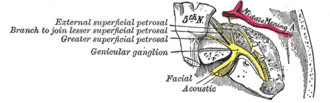
The course and connections of the facial nerve in the temporal bone.
Ear internal anatomy numbered.svg
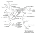
Cranial nerves VII and VIII and selected structures of the inner and middle ear. 1 Nervus vestibularis, 2 Nervus cochlearis, 3 Nervus intermediofacialis, 4 Ganglion geniculi, 5 Chorda tympani, 6 Cochlea, 7 Ductus semicirculares, 8 Malleus, 9 Membrana tympani, 10 Tuba auditiva
Details
Innervates lacrimal glands, submandibular glands, sublingual glands, tongue, palate, pharynx, external auditory meatus, stapedius muscle, posterior belly of the digastric muscle, stylohyoid muscle, muscles of facial expression
Identifiers
Latin ganglion geniculi nervi facialis
MeSH D005830
TA98 A14.2.01.116
TA2 6287
FMA 53414
Anatomical terms of neuroanatomy

The geniculate ganglion (from Latin genu, for "knee" [1] ) is a collection of pseudounipolar sensory neurons of the facial nerve located in the facial canal of the head. It receives fibers from the facial nerve. It sends fibers that supply the lacrimal glands, submandibular glands, sublingual glands, tongue, palate, pharynx, external auditory meatus, stapedius muscle, posterior belly of the digastric muscle, stylohyoid muscle, and muscles of facial expression.

Contents

The geniculate ganglion is one of several ganglia of the head and neck. Like the others, it is a bilaterally distributed structure, with each side of the face having a geniculate ganglion.

Structure

The geniculate ganglion is located close to the internal auditory meatus. [2] It is covered superiorly by the petrous part of the temporal bone (which is sometimes absent over the ganglion). [3]

The geniculate ganglion receives fibers from the motor, sensory, and parasympathetic components of the facial nerve. It contains special sensory neuronal cell bodies for taste, from fibers coming up from the tongue through the chorda tympani and from fibers coming up from the roof of the palate through the greater petrosal nerve. [4] Sensory and parasympathetic inputs are carried into the geniculate ganglion via the nervus intermedius. Motor fibers are carried via the facial nerve proper. The greater petrosal nerve, which carries preganglionic parasympathetic fibers, emerges from the anterior aspect of the ganglion.

The motor fibers of the facial nerve proper and parasympathetic fibers to the submandibular and pterygopalatine ganglia do not synapse in the geniculate ganglion. The afferent fibers carrying pain, temperature, and touch from the posterior auricular nerve, as well as those carrying special sensory (taste) fibers from the tongue (via the chorda tympani), do not synapse in the geniculate ganglion. Instead, the cells of the geniculate ganglion relay the signal to the appropriate brainstem nucleus, much like the Dorsal root ganglion neurons relay signal to nuclei in the spinal cord. [5]

Function

Via the geniculate ganglion, the facial nerve (CN VII) gives [6]

Clinical significance

The geniculate ganglion is an important surgical landmark near the internal auditory meatus. [2]

The geniculate ganglion may become inflamed due to viral infection by herpes zoster virus. [8]

Additional images

See also

Related Research Articles

<span class="mw-page-title-main">Parasympathetic nervous system</span> Division of the autonomic nervous system

The parasympathetic nervous system (PSNS) is one of the three divisions of the autonomic nervous system, the others being the sympathetic nervous system and the enteric nervous system. The enteric nervous system is sometimes considered part of the autonomic nervous system, and sometimes considered an independent system.

<span class="mw-page-title-main">Facial nerve</span> Cranial nerve VII, for the face and tasting

The facial nerve, also known as the seventh cranial nerve, cranial nerve VII, or simply CN VII, is a cranial nerve that emerges from the pons of the brainstem, controls the muscles of facial expression, and functions in the conveyance of taste sensations from the anterior two-thirds of the tongue. The nerve typically travels from the pons through the facial canal in the temporal bone and exits the skull at the stylomastoid foramen. It arises from the brainstem from an area posterior to the cranial nerve VI and anterior to cranial nerve VIII.

Articles related to anatomy include:

<span class="mw-page-title-main">Glossopharyngeal nerve</span> Cranial nerve IX, for the tongue and pharynx

The glossopharyngeal nerve, also known as the ninth cranial nerve, cranial nerve IX, or simply CN IX, is a cranial nerve that exits the brainstem from the sides of the upper medulla, just anterior to the vagus nerve. Being a mixed nerve (sensorimotor), it carries afferent sensory and efferent motor information. The motor division of the glossopharyngeal nerve is derived from the basal plate of the embryonic medulla oblongata, whereas the sensory division originates from the cranial neural crest.

<span class="mw-page-title-main">Mandibular nerve</span> Branch of the trigeminal nerve responsible for the lower face and jaw

In neuroanatomy, the mandibular nerve (V3) is the largest of the three divisions of the trigeminal nerve, the fifth cranial nerve (CN V). Unlike the other divisions of the trigeminal nerve (ophthalmic nerve, maxillary nerve) which contain only afferent fibers, the mandibular nerve contains both afferent and efferent fibers. These nerve fibers innervate structures of the lower jaw and face, such as the tongue, lower lip, and chin. The mandibular nerve also innervates the muscles of mastication.

<span class="mw-page-title-main">Submandibular gland</span> Human salivary gland

The paired submandibular glands are major salivary glands located beneath the floor of the mouth. In adult humans, they each weigh about 15 grams and contribute some 60–67% of unstimulated saliva secretion; on stimulation their contribution decreases in proportion as parotid gland secretion rises to 50%. The average length of the normal adult human submandibular salivary gland is approximately 27 mm, while the average width is approximately 14.3 mm.

<span class="mw-page-title-main">Sublingual gland</span>

The sublingual gland is a seromucous polystomatic exocrine gland. Located underneath the oral diaphragm, the sublingual gland is the smallest and most diffuse of the three major salivary glands of the oral cavity, with the other two being the submandibular and parotid. The sublingual gland provides approximately 3-5% of the total salivary volume.

<span class="mw-page-title-main">Otic ganglion</span>

The otic ganglion is a small parasympathetic ganglion located immediately below the foramen ovale in the infratemporal fossa and on the medial surface of the mandibular nerve. It is functionally associated with the glossopharyngeal nerve and innervates the parotid gland for salivation.

<span class="mw-page-title-main">Pterygopalatine ganglion</span> Parasympathetic ganglion in the pterygopalatine fossa

The pterygopalatine ganglion is a parasympathetic ganglion in the pterygopalatine fossa. It is one of four parasympathetic ganglia of the head and neck,.

<span class="mw-page-title-main">Auriculotemporal nerve</span> Branch of the mandibular nerve

The auriculotemporal nerve is a sensory branch of the mandibular nerve (CN V3) that runs with the superficial temporal artery and vein, and provides sensory innervation to parts of the external ear, scalp, and temporomandibular joint. The nerve also conveys post-ganglionic parasympathetic fibres from the otic ganglion to the parotid gland.

<span class="mw-page-title-main">Chorda tympani</span> Nerve carrying taste sensations

Chorda tympani is a branch of the facial nerve that carries gustatory (taste) sensory innervation from the front of the tongue and parasympathetic (secretomotor) innervation to the submandibular and sublingual salivary glands.

<span class="mw-page-title-main">Greater petrosal nerve</span>

The greater petrosal nerve is a nerve of the head mainly containing pre-ganglionic parasympathetic fibres which ultimately synapse in the pterygopalatine ganglion. It branches from the facial nerve and is derived from the parasympathetic part of the nervus intermedius component of CN VII, with its cell bodies located in the superior salivary nucleus. In the connective tissue substance of the foramen lacerum, the greater petrosal nerve unites with the (sympathetic) deep petrosal nerve to form the nerve of the pterygoid canal which proceeds to the pterygopalatine ganglion.

<span class="mw-page-title-main">Lingual nerve</span> Human nerve relaying sense to the tongue

The lingual nerve carries sensory innervation from the anterior two-thirds of the tongue. It contains fibres from both the mandibular division of the trigeminal nerve (CN V3) and from the facial nerve (CN VII). The fibres from the trigeminal nerve are for touch, pain and temperature (general sensation), and the ones from the facial nerve are for taste (special sensation).

<span class="mw-page-title-main">Facial canal</span> Hole in the temporal bone of the skull carrying the facial nerve

The facial canal is a Z-shaped canal in the temporal bone of the skull. It extends between the internal acoustic meatus and stylomastoid foramen. It transmits the facial nerve.

<span class="mw-page-title-main">Submandibular ganglion</span>

The submandibular ganglion is part of the human autonomic nervous system. It is one of four parasympathetic ganglia of the head and neck..

<span class="mw-page-title-main">Parasympathetic ganglia</span> Autonomic ganglia of the parasympathetic nervous system

Parasympathetic ganglia are the autonomic ganglia of the parasympathetic nervous system. Most are small terminal ganglia or intramural ganglia, so named because they lie near or within (respectively) the organs they innervate. The exceptions are the four paired parasympathetic ganglia of the head and neck.

<span class="mw-page-title-main">Intermediate nerve</span> Portion of the facial nerve

The intermediate nerve, nervus intermedius, nerve of Wrisberg or Glossopalatine nerve, is the part of the facial nerve located between the motor component of the facial nerve and the vestibulocochlear nerve. It contains the sensory and parasympathetic fibers of the facial nerve. Upon reaching the facial canal, it joins with the motor root of the facial nerve at the geniculate ganglion. Alex Alfieri postulates that the intermediate nerve should be considered as a separate cranial nerve and not a part of the facial nerve.

<span class="mw-page-title-main">Salivatory nuclei</span>

The salivatory nuclei are two parasympathetic general visceral efferent cranial nerve nuclei - the superior salivatory nucleus and the inferior salivatory nucleus - that innervate the salivary glands. Both are located in the pontine tegmentum of the brainstem.

<span class="mw-page-title-main">Outline of human anatomy</span> Overview of and topical guide to human anatomy

The following outline is provided as an overview of and topical guide to human anatomy:

References

  1. "genu-, geni-, gen- + (Latin: knee)". WordInfo. Retrieved 2008-10-03.
  2. 1 2 Hall, George M.; Pulec, Jack L.; Rhoton, Albert L. Jr. (1 November 1969). "Geniculate Ganglion Anatomy for the Otologist". Archives of Otolaryngology . 90 (5): 568–571. doi:10.1001/archotol.1969.00770030570007. ISSN   0003-9977.
  3. Rhoton, Albert L.; Pulec, Jack L.; Hall, George M.; Boyd, Allen S. (1 January 1968). "Absence of Bone over the Geniculate Ganglion". Journal of Neurosurgery . 28 (1): 48–53. doi:10.3171/jns.1968.28.1.0048.
  4. M.J. Turlough FitzGerald; Gregory Gruener; Estomih Mtui (2012). Clinical neuroanatomy and neuroscience (6th ed.). Saunders/Elsevier. ISBN   978-0-7020-3738-2.[ page needed ]
  5. Moore, Keith L.; Dalley, Arthur F.; Agur, A. M. R. (2013-02-13). Clinically Oriented Anatomy. Lippincott Williams & Wilkins. ISBN   978-1-4511-1945-9.
  6. Agur., Anne M. R. Clinically Oriented Anatomy, 8e. Lippincott, Williams & Wilkins. ISBN   978-1-4963-4721-3. OCLC   1076750418.
  7. Boudreau, James C.; Sivakumar, Latha; Do, Ly Thi; White, Thomas D.; Oravec, Joseph; Hoang, Nga Kieu (1985). "Neurophysiology of geniculate ganglion (facial nerve) taste systems: species comparisons". Chemical Senses. 10 (1): 89–127. doi:10.1093/chemse/10.1.89. ISSN   0379-864X.
  8. Hunt, J. Ramsay (February 1907). "ON HERPETIC INFLAMMATIONS OF THE GENICULATE GANGLION. A NEW SYNDROME AND ITS COMPLICATIONS". The Journal of Nervous and Mental Disease . 34 (2): 73–96. ISSN   0022-3018.