
Cranial nerves are the nerves that emerge directly from the brain, of which there are conventionally considered twelve pairs. Cranial nerves relay information between the brain and parts of the body, primarily to and from regions of the head and neck, including the special senses of vision, taste, smell, and hearing.

The middle ear is the portion of the ear medial to the eardrum, and distal to the oval window of the cochlea.
The ossicles are three bones in either middle ear that are among the smallest bones in the human body. They serve to transmit sound vibrations sent from the ear drum to the fluid-filled labyrinth (cochlea). The absence of the auditory ossicles would constitute a moderate-to-severe hearing loss. The term "ossicle" literally means "tiny bone". Though the term may refer to any small bone throughout the body, it typically refers to the malleus, incus, and stapes of the middle ear.
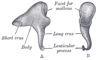
The incus or anvil in the ear is one of three small bones (ossicles) in the middle ear. The incus receives vibrations from the malleus, to which it is connected laterally, and transmits these to the stapes medially. The incus is named for its resemblance to an anvil.

The stapes or stirrup is a bone in the middle ear of humans and other animals which is involved in the conduction of sound vibrations to the inner ear. This bone is connected to the oval window by its annular ligament, which allows the footplate to transmit sound energy through the oval window into the inner ear. The stapes is the smallest and lightest bone in the human body, and is so-called because of its resemblance to a stirrup.

The facial nerve, also known as the seventh cranial nerve, cranial nerve VII, or simply CN VII, is a cranial nerve that emerges from the pons of the brainstem, controls the muscles of facial expression, and functions in the conveyance of taste sensations from the anterior two-thirds of the tongue. The nerve typically travels from the pons through the facial canal in the temporal bone and exits the skull at the stylomastoid foramen. It arises from the brainstem from an area posterior to the cranial nerve VI and anterior to cranial nerve VIII.
Articles related to anatomy include:

An ear is the organ that enables hearing and body balance using the vestibular system. In mammals, the ear is usually described as having three parts: the outer ear, the middle ear and the inner ear. The outer ear consists of the pinna and the ear canal. Since the outer ear is the only visible portion of the ear in most animals, the word "ear" often refers to the external part alone. The middle ear includes the tympanic cavity and the three ossicles. The inner ear sits in the bony labyrinth, and contains structures which are key to several senses: the semicircular canals, which enable balance and eye tracking when moving; the utricle and saccule, which enable balance when stationary; and the cochlea, which enables hearing. The ear canal is cleaned via earwax, which naturally migrates to the auricle. The ears of vertebrates are placed somewhat symmetrically on either side of the head, an arrangement that aids sound localization.

The acoustic reflex is an involuntary muscle contraction that occurs in the middle ear in response to loud sound stimuli or when the person starts to vocalize.

The tensor tympani is a muscle within the middle ear, located in the bony canal above the bony part of the auditory tube, and connects to the malleus bone. Its role is to dampen loud sounds, such as those produced from chewing, shouting, or thunder. Because its reaction time is not fast enough, the muscle cannot protect against hearing damage caused by sudden loud sounds, like explosions or gunshots.

The tympanic cavity is a small cavity surrounding the bones of the middle ear. Within it sit the ossicles, three small bones that transmit vibrations used in the detection of sound.

The pharyngeal arches, also known as visceral arches, are structures seen in the embryonic development of vertebrates that are recognisable precursors for many structures. In fish, the arches are known as the branchial arches, or gill arches.

The marginal mandibular branch of the facial nerve arises from the facial nerve in the parotid gland at the parotid plexus. It passes anterior-ward deep to the platysma and depressor anguli oris muscles. It provides motor innervation to muscles of the lower lip and chin: the depressor labii inferioris muscle, depressor anguli oris muscle, and mentalis muscle. It communicates with the mental branch of the inferior alveolar nerve.
The posterior auricular nerve is a nerve of the head. It is a branch of the facial nerve. It communicates with branches from the vagus nerve, the great auricular nerve, and the lesser occipital nerve. Its auricular branch supplies the posterior auricular muscle, the intrinsic muscles of the auricle, and gives sensation to the auricle. Its occipital branch supplies the occipitalis muscle.

The pyramidal eminence is a hollow conical projection upon the posterior wall of the tympanic cavity of the middle ear. The stapedius muscle arises in the hollow of the eminence and its tendon exits through its apex.
The nerve to the stapedius is a branch of the facial nerve which innervates the stapedius muscle. It arises from the CN VII within the facial canal, opposite the pyramidal eminence. It passes through a small canal in this eminence to reach the stapedius muscle.

The facial muscles are a group of striated skeletal muscles supplied by the facial nerve that, among other things, control facial expression. These muscles are also called mimetic muscles. They are only found in mammals, although they derive from neural crest cells found in all vertebrates. They are the only muscles that attach to the dermis.

The following outline is provided as an overview of and topical guide to human anatomy:

The anterior auricular muscle, the smallest of the three auricular muscles, is thin and fan-shaped, and its fibers are pale and indistinct. It arises from the lateral edge of the epicranial aponeurosis, and its fibers converge to be inserted into a projection on the front of the helix.
Neurotology or neuro-otology is a subspecialty of otolaryngology—head and neck surgery, also known as ENT medicine. Neuro-otology is closely related to otology, clinical neurology and neurosurgery.
Rodríguez-Vázquez JF. Development of the stapedius muscle and pyramidal eminence in humans. J Anat. 2009 Sep;215(3):292-9. doi: 10.1111/j.1469-7580.2009.01105.x

