The middle ear is the portion of the ear medial to the eardrum, and distal to the oval window of the cochlea.

The facial nerve, also known as the seventh cranial nerve, cranial nerve VII, or simply CN VII, is a cranial nerve that emerges from the pons of the brainstem, controls the muscles of facial expression, and functions in the conveyance of taste sensations from the anterior two-thirds of the tongue. The nerve typically travels from the pons through the facial canal in the temporal bone and exits the skull at the stylomastoid foramen. It arises from the brainstem from an area posterior to the cranial nerve VI and anterior to cranial nerve VIII.
Articles related to anatomy include:

In anatomy, the palatine bones are two irregular bones of the facial skeleton in many animal species, located above the uvula in the throat. Together with the maxilla, they comprise the hard palate.

In anatomy, the orbit is the cavity or socket/hole of the skull in which the eye and its appendages are situated. "Orbit" can refer to the bony socket, or it can also be used to imply the contents. In the adult human, the volume of the orbit is 30 millilitres, of which the eye occupies 6.5 ml. The orbital contents comprise the eye, the orbital and retrobulbar fascia, extraocular muscles, cranial nerves II, III, IV, V, and VI, blood vessels, fat, the lacrimal gland with its sac and duct, the eyelids, medial and lateral palpebral ligaments, cheek ligaments, the suspensory ligament, septum, ciliary ganglion and short ciliary nerves.

The fourth ventricle is one of the four connected fluid-filled cavities within the human brain. These cavities, known collectively as the ventricular system, consist of the left and right lateral ventricles, the third ventricle, and the fourth ventricle. The fourth ventricle extends from the cerebral aqueduct to the obex, and is filled with cerebrospinal fluid (CSF).

The posterior cranial fossa is the part of the cranial cavity located between the foramen magnum, and tentorium cerebelli. It is formed by the sphenoid bones, temporal bones, and occipital bone. It lodges the cerebellum, and parts of the brainstem.

The stapedius is the smallest skeletal muscle in the human body. At just over one millimeter in length, its purpose is to stabilize the smallest bone in the body, the stapes or stirrup bone of the middle ear.

The tympanic cavity is a small cavity surrounding the bones of the middle ear. Within it sit the ossicles, three small bones that transmit vibrations used in the detection of sound.

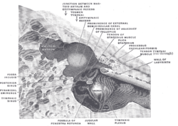
The greater petrosal nerve is a nerve of the head mainly containing pre-ganglionic parasympathetic fibres which ultimately synapse in the pterygopalatine ganglion. It branches from the facial nerve and is derived from the parasympathetic part of the nervus intermedius component of CN VII, with its cell bodies located in the superior salivary nucleus. In the connective tissue substance of the foramen lacerum, the greater petrosal nerve unites with the (sympathetic) deep petrosal nerve to form the nerve of the pterygoid canal which proceeds to the pterygopalatine ganglion.

The internal auditory meatus is a canal within the petrous part of the temporal bone of the skull between the posterior cranial fossa and the inner ear.

The mastoid part of the temporal bone is the posterior (back) part of the temporal bone, one of the bones of the skull. Its rough surface gives attachment to various muscles and it has openings for blood vessels. From its borders, the mastoid part articulates with two other bones.

The petrous part of the temporal bone is pyramid-shaped and is wedged in at the base of the skull between the sphenoid and occipital bones. Directed medially, forward, and a little upward, it presents a base, an apex, three surfaces, and three angles, and houses in its interior, the components of the inner ear. The petrous portion is among the most basal elements of the skull and forms part of the endocranium. Petrous comes from the Latin word petrosus, meaning "stone-like, hard". It is one of the densest bones in the body. In other mammals, it is a separate bone, the petrosal bone.

The facial canal is a Z-shaped canal in the temporal bone of the skull. It extends between the internal acoustic meatus and stylomastoid foramen. It transmits the facial nerve.
The nerve to the stapedius is a branch of the facial nerve which innervates the stapedius muscle. It arises from the CN VII within the facial canal, opposite the pyramidal eminence. It passes through a small canal in this eminence to reach the stapedius muscle.

The aditus to mastoid antrum is a large, irregular opening upon the posterior wall of the tympanic cavity by which the mastoid antrum communicates with the epitympanic recess of the tympanic cavity. The walls of the antrum are lined by mucosa which is continuous with that lining the mastoid cells and tympanic cavity.

The promontory of the tympanic cavity, also known as the cochlear promontory is a rounded hollow prominence upon - and most prominent feature of - the medial wall of the tympanic cavity formed by the underlying first turn of the cochlea. The surface of the promontory is furrowed by fine grooves that accommodate to the strands of the tympanic plexus.

The vestibule is the central part of the bony labyrinth in the inner ear, and is situated medial to the eardrum, behind the cochlea, and in front of the three semicircular canals.

The following outline is provided as an overview of and topical guide to human anatomy:
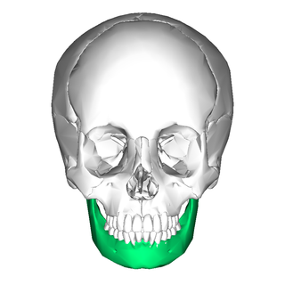
In jawed vertebrates, the mandible, lower jaw, or jawbone is a bone that makes up the lower – and typically more mobile – component of the mouth.