Torus tubarius

Last updated
Torus tubarius
Gray915.png
Auditory tube, laid open by a cut in its long axis (torus tubarius not labeled)
Details
Identifiers
Latin Torus tubarius
TA A05.3.01.012
FMA 54993
Anatomical terminology

The base of the cartilaginous portion of the auditory tube (eustachian tube, pharyngotympanic tube) lies directly under the mucous membrane of the nasal part of the pharynx, where it forms an elevation, the torus tubarius, the torus of the auditory tube, or cushion, behind the pharyngeal orifice of the tube. The torus tubarius is very close to the tubal tonsil, [1] which is sometimes also called the tonsil of (the) torus tubarius. [2] Equating the torus with its tonsil however might be seen as incorrect or imprecise.

Cartilage resilient and smooth elastic tissue, rubber-like padding that covers and protects the ends of long bones at the joints

Cartilage is a resilient and smooth elastic tissue, a rubber-like padding that covers and protects the ends of long bones at the joints, and is a structural component of the rib cage, the ear, the nose, the bronchial tubes, the intervertebral discs, and many other body components. It is not as hard and rigid as bone, but it is much stiffer and much less flexible than muscle. The matrix of cartilage is made up of glycosaminoglycans, proteoglycans,collagen fibers and, sometimes, elastin.

Mucous membrane the protective layer, which lines the interior of hollow organs

A mucous membrane or mucosa is a membrane that lines various cavities in the body and covers the surface of internal organs. It consists of one or more layers of epithelial cells overlying a layer of loose connective tissue. It is mostly of endodermal origin and is continuous with the skin at various body openings such as the eyes, ears, inside the nose, inside the mouth, lip, vagina, the urethral opening and the anus. Some mucous membranes secrete mucus, a thick protective fluid. The function of the membrane is to stop pathogens and dirt from entering the body and to prevent bodily tissues from becoming dehydrated.

Tubal tonsil

The tubal tonsil is one of the four main tonsil groups comprising Waldeyer's tonsillar ring, which also includes the palatine tonsils, the lingual tonsils, and the pharyngeal tonsils.

Two folds run posteriorly and anteriorly:
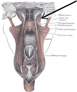
Salpingopharyngeus muscle

The salpingopharyngeus muscle arises from the superior border of the medial cartilage of the pharyngotympanic tube, in the nasal cavity, making the posterior welt of the torus tubarius; it passes downward and blends with the posterior fasciculus of the palatopharyngeus muscle.

Palatopharyngeus muscle

The palatopharyngeusmuscle is a small muscle in the roof of the mouth.

Related Research Articles

Larynx voice box, an organ in the neck of amphibians, reptiles, and mammals

The larynx, commonly called the voice box, is an organ in the top of the neck of tetrapods involved in breathing, producing sound, and protecting the trachea against food aspiration. The larynx houses the vocal folds, and manipulates pitch and volume, which is essential for phonation. It is situated just below where the tract of the pharynx splits into the trachea and the esophagus. The word larynx comes from a similar Ancient Greek word.

Articles related to anatomy include:

Eustachian tube

The Eustachian tube, also known as the auditory tube or pharyngotympanic tube, is a tube that links the nasopharynx to the middle ear. It is a part of the middle ear. In adult humans the Eustachian tube is approximately 35 mm (1.4 in) long and 3 mm (0.12 in) in diameter. It is named after the sixteenth-century Italian anatomist Bartolomeo Eustachi.

Inferior nasal concha facial bone

The inferior nasal concha is one of the three paired nasal conchae in the nose. It extends horizontally along the lateral wall of the nasal cavity and consists of a lamina of spongy bone, curled upon itself like a scroll,. The inferior nasal conchae are considered a pair of facial bones. As the air passes through the turbinates, the air is churned against these mucosa-lined bones in order to receive warmth, moisture and cleansing. Superior to inferior nasal concha are the middle nasal concha and superior nasal concha which arise from the cranial portion of the skull. Hence, these two are considered as a part of the cranial bones.

Pterygopalatine ganglion

The pterygopalatine ganglion is a parasympathetic ganglion found in the pterygopalatine fossa. It is largely innervated by the greater petrosal nerve ; and its axons project to the lacrimal glands and nasal mucosa. The flow of blood to the nasal mucosa, in particular the venous plexus of the conchae, is regulated by the pterygopalatine ganglion and heats or cools the air in the nose. It is one of four parasympathetic ganglia of the head and neck, the others being the submandibular ganglion, otic ganglion, and ciliary ganglion.

Arytenoid cartilage

The arytenoid cartilages are a pair of small three-sided pyramids which form part of the larynx, to which the vocal folds are attached. These allow and aid in the vocal cords' movement.

Stylopharyngeus muscle

The stylopharyngeus is a muscle in the head that stretches between the temporal styloid process and the pharynx.

Tensor veli palatini muscle

The tensor veli palatini muscle is a broad, thin, ribbon-like muscle in the head that tenses the soft palate.

Nasociliary nerve

The nasociliary nerve is a branch of the ophthalmic nerve (CN V1; one of three branches of the trigeminal nerve a.k.a. CN V). It is intermediate in size between the two other main branches of the ophthalmic nerve, the frontal nerve and the lacrimal nerve, and is more deeply placed.

Pharyngeal arch

The pharyngeal arches —also known as visceral archesare structures seen in the embryonic development of vertebrates that are recognisable precursors for many structures. In fish the arches are known as the branchial arches or gill arches.

Laryngeal vestibule

The portion of the cavity of the larynx above the vocal folds is called the laryngeal vestibule; it is wide and triangular in shape, its base or anterior wall presenting, however, about its center the backward projection of the tubercle of the epiglottis. It contains the vestibular folds, and between these and the vocal folds are the laryngeal ventricles.

Aryepiglottic fold Folds near the larynx

The Aryepiglottic folds are triangular folds of mucous membrane enclosing ligamentous and muscular fibres. They are located at the entrance of the larynx, extending from the lateral borders of the epiglottis to the arytenoid cartilages, hence the name 'aryepiglottic'. They contain the aryepiglottic muscles and form the upper borders of the quadrangular membrane.

Human nose feature of the face

The human nose is the most protruding part of the face that bears the nostrils and is the first organ of the respiratory system. The nose is also the principal organ in the olfactory system. The shape of the nose is determined by the nasal bones and the nasal cartilages, including the nasal septum which separates the nostrils and divides the nasal cavity into two. On average the nose of a male is larger than that of a female.

Laryngeal ventricle

The laryngeal ventricle, is a fusiform fossa, situated between the vestibular and vocal folds on either side, and extending nearly their entire length. There is also a sinus of Morgagni in the pharynx.

Outline of human anatomy scientific study of the morphology of the human body

The following outline is provided as an overview of and topical guide to human anatomy:

Pharynx part of the throat that is behind the mouth and nasal cavity

The pharynx is the part of the throat behind the mouth and nasal cavity and above the esophagus and larynx, or the tubes going down to the stomach and the lungs. It is found in vertebrates and invertebrates, though its structure varies across species.

Histology is the study of the minute structure, composition, and function of tissues. Mature human vocal cords are composed of layered structures which are quite different at the histological level.

References

This article incorporates text in the public domain from page 1043 of the 20th edition of Gray's Anatomy (1918)

The public domain consists of all the creative works to which no exclusive intellectual property rights apply. Those rights may have expired, been forfeited, expressly waived, or may be inapplicable.

<i>Grays Anatomy</i> English-language textbook of human anatomy

Gray's Anatomy is an English language textbook of human anatomy originally written by Henry Gray and illustrated by Henry Vandyke Carter. Earlier editions were called Anatomy: Descriptive and Surgical and Gray's Anatomy: Descriptive and Applied, but the book's name is commonly shortened to, and later editions are titled, Gray's Anatomy. The book is widely regarded as an extremely influential work on the subject, and has continued to be revised and republished from its initial publication in 1858 to the present day. The latest edition of the book, the 41st, was published in September 2015.

Loyola University Chicago university

Loyola University Chicago is a private Catholic research university in Chicago, Illinois. Founded in 1870 by the Jesuits, today Loyola is one of the largest Catholic universities in the United States. Loyola's professional schools have educated generations of local business and civic leaders, and distinguished programs in medicine, nursing, and health sciences are anchored by the nationally recognized Loyola University Medical Center.