Paranasal sinuses

Last updated
Paranasal sinuses
Paranasal Sinuses ant.jpg
Paranasal sinuses seen in a frontal view
Paranasal Sinuses lat.jpg
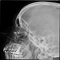
Lateral projection of the paranasal sinuses
Details
Identifiers
Latin sinus paranasales
MeSH D010256
TA98 A06.1.03.001
TA2 3176
FMA 59679
Anatomical terminology

Paranasal sinuses are a group of four paired air-filled spaces that surround the nasal cavity. [1] The maxillary sinuses are located under the eyes; the frontal sinuses are above the eyes; the ethmoidal sinuses are between the eyes and the sphenoidal sinuses are behind the eyes. The sinuses are named for the facial bones and sphenoid bone in which they are located. Their role is disputed.

Contents

Structure

Humans possess four pairs of paranasal sinuses, divided into subgroups that are named according to the bones within which the sinuses lie. They are all innervated by branches of the trigeminal nerve (CN V).

The paranasal sinuses are lined with respiratory epithelium (ciliated pseudostratified columnar epithelium).

Functions

One known function of the paranasal sinuses is the production of nitric oxide, which also functions as a facilitator of oxygen uptake. [3]

Development

Paranasal sinuses form developmentally through excavation of bone by air-filled sacs (pneumatic diverticula) from the nasal cavity. This process begins prenatally (intrauterine life), and it continues through the course of an organism's lifetime.

The results of experimental studies suggest that the natural ventilation rate of a sinus with a single sinus ostium (opening) is extremely slow. Such limited ventilation may be protective for the sinus, as it would help prevent drying of its mucosal surface and maintain a near-sterile environment with high carbon dioxide concentrations and minimal pathogen access. Thus composition of gas content in the maxillary sinus is similar to venous blood, with high carbon dioxide and lower oxygen levels compared to breathing air. [4]

At birth, only the maxillary sinus and the ethmoid sinus are developed but not yet pneumatized; only by the age of seven they are fully aerated. The sphenoid sinus appears at the age of three, and the frontal sinuses first appear at the age of six, and fully develop during adulthood. [5]

CT scans, radiographs (x-ray) and other illustrations

Clinical significance

Inflammation

The paranasal sinuses are joined to the nasal cavity via small orifices called ostia. These become blocked easily by allergic inflammation, or by swelling in the nasal lining that occurs with a cold. If this happens, normal drainage of mucus within the sinuses is disrupted, and sinusitis may occur. Because the maxillary posterior teeth are close to the maxillary sinus, this can also cause clinical problems if any disease processes are present, such as an infection in any of these teeth. These clinical problems can include secondary sinusitis, the inflammation of the sinuses from another source such as an infection of the adjacent teeth. [6]

These conditions may be treated with drugs such as decongestants, which cause vasoconstriction in the sinuses; reducing inflammation; by traditional techniques of nasal irrigation; or by corticosteroid.[ medical citation needed ]

Cancer

Malignancies of the paranasal sinuses comprise approximately 0.2% of all malignancies. About 80% of these malignancies arise in the maxillary sinus. Men are much more often affected than women. They most often occur in the age group between 40 and 70 years. Carcinomas are more frequent than sarcomas. Metastases are rare. Tumours of the sphenoid and frontal sinuses are extremely rare.

Etymology

Sinus is a Latin word meaning a fold, curve, or bay. Compare sine .

Other animals

Paranasal sinuses occur in many other animals, including most mammals, birds, non-avian dinosaurs, and crocodilians. The bones occupied by sinuses are quite variable in these other species.

Illustrations

See also

Related Research Articles

Sinus may refer to:

<span class="mw-page-title-main">Ethmoid bone</span> Bone of the facial skeletonl

The ethmoid bone is an unpaired bone in the skull that separates the nasal cavity from the brain. It is located at the roof of the nose, between the two orbits. The cubical bone is lightweight due to a spongy construction. The ethmoid bone is one of the bones that make up the orbit of the eye.

<span class="mw-page-title-main">Sphenoid bone</span> Bone of the neurocranium

The sphenoid bone is an unpaired bone of the neurocranium. It is situated in the middle of the skull towards the front, in front of the basilar part of the occipital bone. The sphenoid bone is one of the seven bones that articulate to form the orbit. Its shape somewhat resembles that of a butterfly or bat with its wings extended.

<span class="mw-page-title-main">Nasal cavity</span> Large, air-filled space above and behind the nose in the middle of the face

The nasal cavity is a large, air-filled space above and behind the nose in the middle of the face. The nasal septum divides the cavity into two cavities, also known as fossae. Each cavity is the continuation of one of the two nostrils. The nasal cavity is the uppermost part of the respiratory system and provides the nasal passage for inhaled air from the nostrils to the nasopharynx and rest of the respiratory tract.

<span class="mw-page-title-main">Orbit (anatomy)</span> Cavity or socket of the skull in which the eye and its appendages are situated

In anatomy, the orbit is the cavity or socket/hole of the skull in which the eye and its appendages are situated. "Orbit" can refer to the bony socket, or it can also be used to imply the contents. In the adult human, the volume of the orbit is 30 millilitres, of which the eye occupies 6.5 ml. The orbital contents comprise the eye, the orbital and retrobulbar fascia, extraocular muscles, cranial nerves II, III, IV, V, and VI, blood vessels, fat, the lacrimal gland with its sac and duct, the eyelids, medial and lateral palpebral ligaments, cheek ligaments, the suspensory ligament, septum, ciliary ganglion and short ciliary nerves.

<span class="mw-page-title-main">Maxillary sinus</span> Largest of the paranasal sinuses, and drains into the middle meatus of the nose

The pyramid-shaped maxillary sinus is the largest of the paranasal sinuses, located in the maxilla. It drains into the middle meatus of the nose through the semilunar hiatus. It is located to the side of the nasal cavity, and below the orbit.

<span class="mw-page-title-main">Ethmoid sinus</span> Air-filled space near the nasal cavity

The ethmoid sinuses or ethmoid air cells of the ethmoid bone are one of the four paired paranasal sinuses. Unlike the other three pairs of paranasal sinuses which consist of one or two large cavities, the ethmoidal sinuses entail a number of small air-filled cavities. The cells are located within the lateral mass (labyrinth) of each ethmoid bone and are variable in both size and number. The cells are grouped into anterior, middle, and posterior groups; the groups differ in their drainage modalities, though all ultimately drain into either the superior or the middle nasal meatus of the lateral wall of the nasal cavity.

<span class="mw-page-title-main">Ophthalmic nerve</span> Sensory nerve of the face

The ophthalmic nerve (CN V1) is a sensory nerve of the head. It is one of three divisions of the trigeminal nerve (CN V), a cranial nerve. It has three major branches which provide sensory innervation to the eye, and the skin of the upper face and anterior scalp, as well as other structures of the head.

<span class="mw-page-title-main">Nasociliary nerve</span> Branch of the ophthalmic nerve

The nasociliary nerve is a branch of the ophthalmic nerve (CN V1) (which is in turn a branch of the trigeminal nerve (CN V)). It is intermediate in size between the other two branches of the ophthalmic nerve, the frontal nerve and lacrimal nerve.

<span class="mw-page-title-main">Sphenoid sinus</span> One of the four paired paranasal sinuses

The sphenoid sinus is a paired paranasal sinus occurring within the body of the sphenoid bone. It represents one pair of the four paired paranasal sinuses. The pair of sphenoid sinuses are separated in the middle by a septum of sphenoid sinuses. Each sphenoid sinus communicates with the nasal cavity via the opening of sphenoidal sinus. The two sphenoid sinuses vary in size and shape, and are usually asymmetrical.

<span class="mw-page-title-main">Ethmoidal labyrinth</span>

The ethmoidal labyrinth or lateral mass of the ethmoid bone consists of a number of thin-walled cellular cavities, the ethmoid air cells, arranged in three groups, anterior, middle, and posterior, and interposed between two vertical plates of bone; the lateral plate forms part of the orbit, the medial plate forms part of the nasal cavity. In the disarticulated bone many of these cells are opened into, but when the bones are articulated, they are closed in at every part, except where they open into the nasal cavity.

<span class="mw-page-title-main">Anterior ethmoidal artery</span>

The anterior ethmoidal artery is a branch of the ophthalmic artery in the orbit. It exits the orbit through the anterior ethmoidal foramen alongside the anterior ethmoidal nerve. It contributes blood supply to the ethmoid sinuses, frontal sinuses, the dura mater, lateral nasal wall, and nasal septum. It issues a meningeal branch, and nasal branches.

<span class="mw-page-title-main">Anterior ethmoidal nerve</span> Nerve of the nose

The anterior ethmoidal nerve is a nerve of the head. It is a branch of the nasociliary nerve (itself a branch of the ophthalmic nerve (V1)). It arises in the orbit, and enters first the cranial cavity and then the nasal cavity. It provides sensory innervation to part of the meninges, parts of the nasal cavity, and part of the skin of the nose.

<span class="mw-page-title-main">Anterior cranial fossa</span>

The anterior cranial fossa is a depression in the floor of the cranial base which houses the projecting frontal lobes of the brain. It is formed by the orbital plates of the frontal, the cribriform plate of the ethmoid, and the small wings and front part of the body of the sphenoid; it is limited behind by the posterior borders of the small wings of the sphenoid and by the anterior margin of the chiasmatic groove. The lesser wings of the sphenoid separate the anterior and middle fossae.

<span class="mw-page-title-main">Posterior ethmoidal nerve</span> Nerve of the orbit around the eye

The posterior ethmoidal nerve is a nerve of the head. It is a branch of the nasociliary nerve (itself a branch of the ophthalmic nerve (CN V1)). It provides sensory innervation to the sphenoid sinus and ethmoid sinus, and part of the dura mater in the anterior cranial fossa.

<span class="mw-page-title-main">Body of sphenoid bone</span>

The body of the sphenoid bone, more or less cubical in shape, is hollowed out in its interior to form two large cavities, the sphenoidal sinuses, which are separated from each other by a septum.

<span class="mw-page-title-main">Human nose</span> Feature of the human face

The human nose is the first organ of the respiratory system. It is also the principal organ in the olfactory system. The shape of the nose is determined by the nasal bones and the nasal cartilages, including the nasal septum, which separates the nostrils and divides the nasal cavity into two.

A sinus is a sac or cavity in any organ or tissue, or an abnormal cavity or passage. In common usage, "sinus" usually refers to the paranasal sinuses, which are air cavities in the cranial bones, especially those near the nose and connecting to it. Most individuals have four paired cavities located in the cranial bone or skull.

<span class="mw-page-title-main">Nasal mucosa</span> Part of the mucus membrane lining the nasal cavity

The nasal mucosa lines the nasal cavity. It is part of the respiratory mucosa, the mucous membrane lining the respiratory tract. The nasal mucosa is intimately adherent to the periosteum or perichondrium of the nasal conchae. It is continuous with the skin through the nostrils, and with the mucous membrane of the nasal part of the pharynx through the choanae. From the nasal cavity its continuity with the conjunctiva may be traced, through the nasolacrimal and lacrimal ducts; and with the frontal, ethmoidal, sphenoidal, and maxillary sinuses, through the several openings in the nasal meatuses. The mucous membrane is thickest, and most vascular, over the nasal conchae. It is also thick over the nasal septum where increased numbers of goblet cells produce a greater amount of nasal mucus. It is very thin in the meatuses on the floor of the nasal cavities, and in the various sinuses. It is one of the most commonly infected tissues in adults and children. Inflammation of this tissue may cause significant impairment of daily activities, with symptoms such as stuffy nose, headache, mouth breathing, etc.

<span class="mw-page-title-main">Nasal meatus</span> Nasal passage of the nasal cavity

In anatomy, the term nasal meatus can refer to any of the three meatuses (passages) through the skull's nasal cavity: the superior meatus, middle meatus, and inferior meatus.

References

  1. "Paranasal sinuses". 23 December 2021.
  2. 1 2 3 "Paranasal Sinus Anatomy: Overview, Gross Anatomy, Microscopic Anatomy". 2016-08-24.
  3. Lundberg, Jon O (November 2008). "Nitric oxide and the paranasal sinuses". The Anatomical Record: Advances in Integrative Anatomy and Evolutionary Biology. 291 (11): 1479–1484. doi: 10.1002/ar.20782 . PMID   18951492.
  4. "ARTICLES | Journal of Applied Physiology". jap.physiology.org. Retrieved 2017-09-07.
  5. Towbin, Richard; Dunbar, J. Scott (1982). "The paranasal sinuses in childhood". RadioGraphics. 2 (2): 253–279. doi: 10.1148/radiographics.2.2.253 .
  6. Illustrated Anatomy of the Head and Neck, Fehrenbach and Herring, Elsevier, 2012, p. 68