Articles related to anatomy include:

Rhinoplasty, commonly called nose job, medically called nasal reconstruction is a plastic surgery procedure for altering and reconstructing the nose. There are two types of plastic surgery used – reconstructive surgery that restores the form and functions of the nose and cosmetic surgery that changes the appearance of the nose. Reconstructive surgery seeks to resolve nasal injuries caused by various traumas including blunt, and penetrating trauma and trauma caused by blast injury. Reconstructive surgery can also treat birth defects, breathing problems, and failed primary rhinoplasties. Rhinoplasty may remove a bump, narrow nostril width, change the angle between the nose and the mouth, or address injuries, birth defects, or other problems that affect breathing, such as a deviated nasal septum or a sinus condition. Surgery only on the septum is called a septoplasty.

The sphenoid bone is an unpaired bone of the neurocranium. It is situated in the middle of the skull towards the front, in front of the basilar part of the occipital bone. The sphenoid bone is one of the seven bones that articulate to form the orbit. Its shape somewhat resembles that of a butterfly or bat with its wings extended.

The inferior nasal concha is one of the three paired nasal conchae in the nose. It extends horizontally along the lateral wall of the nasal cavity and consists of a lamina of spongy bone, curled upon itself like a scroll,. The inferior nasal conchae are considered a pair of facial bones. As the air passes through the turbinates, the air is churned against these mucosa-lined bones in order to receive warmth, moisture and cleansing. Superior to inferior nasal concha are the middle nasal concha and superior nasal concha which both arise from the ethmoid bone, of the cranial portion of the skull. Hence, these two are considered as a part of the cranial bones.

The vomer is one of the unpaired facial bones of the skull. It is located in the midsagittal line, and articulates with the sphenoid, the ethmoid, the left and right palatine bones, and the left and right maxillary bones. The vomer forms the inferior part of the nasal septum in humans, with the superior part formed by the perpendicular plate of the ethmoid bone. The name is derived from the Latin word for a ploughshare and the shape of the bone.

In anatomy, the axis or epistropheus is the second cervical vertebra (C2) of the spine, immediately inferior to the atlas, upon which the head rests.

The nasal cavity is a large, air-filled space above and behind the nose in the middle of the face. The nasal septum divides the cavity into two cavities, also known as fossae. Each cavity is the continuation of one of the two nostrils. The nasal cavity is the uppermost part of the respiratory system and provides the nasal passage for inhaled air from the nostrils to the nasopharynx and rest of the respiratory tract.

Septoplasty [ˈsɛp.toˌplæ.sti] (Etymology: L, saeptum, septum; Gk, πλάσσειν plassein – to shape), or alternatively submucous septal resection and septal reconstruction, is a corrective surgical procedure done to straighten a deviated nasal septum – the nasal septum being the partition between the two nasal cavities. Ideally, the septum should run down the center of the nose. When it deviates into one of the cavities, it narrows that cavity and impedes airflow. Deviated nasal septum or “crooked” internal nose can occur at childbirth or as the result of an injury or other trauma. If the wall that functions as a separator of both sides of the nose is tilted towards one side at a degree greater than 50%, it might cause difficulty breathing. Often the inferior turbinate on the opposite side enlarges, which is termed compensatory hypertrophy. Deviations of the septum can lead to nasal obstruction. Most surgeries are completed in 60 minutes or less, while the recovery time could be up to several weeks. Put simply, septoplasty is a surgery that helps repair the passageways in the nose making it easier to breathe. This surgery is usually performed on patients with a deviated septum, recurrent rhinitis, or sinus issues.

The nasal septum separates the left and right airways of the nasal cavity, dividing the two nostrils.

The greater wing of the sphenoid bone, or alisphenoid, is a bony process of the sphenoid bone; there is one on each side, extending from the side of the body of the sphenoid and curving upward, laterally, and backward.
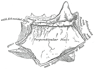
The perpendicular plate of the ethmoid bone is a thin, flattened lamina, polygonal in form, which descends from the under surface of the cribriform plate, and assists in forming the septum of the nose; it is generally deflected a little to one or other side. The anterior border articulates with the spine of the frontal bone and the crest of the nasal bones.

The horizontal plate of palatine bone is a quadrilateral part of the palatine bone, and has two surfaces and four borders.
The piriform aperture, pyriform aperture, or anterior nasal aperture, is a pear-shaped opening in the human skull. Its long axis is vertical, and narrow end upward; in the recent state it is much contracted by the lateral nasal cartilage and the greater and lesser alar cartilages of the nose.
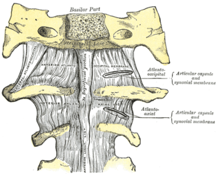
The atlanto-axial joint is a joint in the upper part of the neck between the atlas bone and the axis bone, which are the first and second cervical vertebrae. It is a pivot joint.
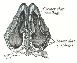
The major alar cartilage is a thin, flexible plate, situated immediately below the lateral nasal cartilage, and bent upon itself in such a manner as to form the medial wall and lateral wall of the nostril of its own side.
The septal nasal cartilage is composed of hyaline cartilage. It is somewhat quadrilateral in form, thicker at its margins than at its center, and completes the separation between the nasal cavities in front.
The nasal cartilages are structures within the nose that provide form and support to the nasal cavity. The nasal cartilages are made up of a flexible material called hyaline cartilage in the distal portion of the nose. There are five individual cartilages that make up the nasal cavity: septal nasal cartilage, lateral nasal cartilage, major alar cartilage, minor alar cartilage, and vomeronasal cartilage.

The articular capsule of the knee joint is the wide and lax joint capsule of the knee. It is thin in front and at the side, and contains the patella, ligaments, menisci, and bursae of the knee. The capsule consists of an inner synovial membrane, and an outer fibrous membrane separated by fatty deposits anteriorly and posteriorly.

The human nose is the most protruding part of the face. It bears the nostrils and is the first organ of the respiratory system. It is also the principal organ in the olfactory system. The shape of the nose is determined by the nasal bones and the nasal cartilages, including the nasal septum which separates the nostrils and divides the nasal cavity into two. On average the nose of a male is larger than that of a female.

The pelvis is the lower part of the trunk, between the abdomen and the thighs, together with its embedded skeleton.
