Paranasal sinuses are a group of four paired air-filled spaces that surround the nasal cavity. The maxillary sinuses are located under the eyes; the frontal sinuses are above the eyes; the ethmoidal sinuses are between the eyes and the sphenoidal sinuses are behind the eyes. The sinuses are named for the facial bones and sphenoid bone in which they are located. Their role is disputed.

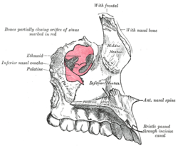
The ethmoid bone is an unpaired bone in the skull that separates the nasal cavity from the brain. It is located at the roof of the nose, between the two orbits. The cubical bone is lightweight due to a spongy construction. The ethmoid bone is one of the bones that make up the orbit of the eye.

The inferior nasal concha is one of the three paired nasal conchae in the nose. It extends horizontally along the lateral wall of the nasal cavity and consists of a lamina of spongy bone, curled upon itself like a scroll,. The inferior nasal conchae are considered a pair of facial bones. As the air passes through the turbinates, the air is churned against these mucosa-lined bones in order to receive warmth, moisture and cleansing. Superior to inferior nasal concha are the middle nasal concha and superior nasal concha which both arise from the ethmoid bone, of the cranial portion of the skull. Hence, these two are considered as a part of the cranial bones.

The nasal cavity is a large, air-filled space above and behind the nose in the middle of the face. The nasal septum divides the cavity into two cavities, also known as fossae. Each cavity is the continuation of one of the two nostrils. The nasal cavity is the uppermost part of the respiratory system and provides the nasal passage for inhaled air from the nostrils to the nasopharynx and rest of the respiratory tract.

In anatomy, a process is a projection or outgrowth of tissue from a larger body. For instance, in a vertebra, a process may serve for muscle attachment and leverage, or to fit, with another vertebra. The word is also used at the microanatomic level, where cells can have processes such as cilia or pedicels. Depending on the tissue, processes may also be called by other terms, such as apophysis, tubercle, or protuberance.

The pyramid-shaped maxillary sinus is the largest of the paranasal sinuses, located in the maxilla. It drains into the middle meatus of the nose through the semilunar hiatus. It is located to the side of the nasal cavity, and below the orbit.

The orbital or horizontal part of the frontal bone consists of two thin triangular plates, the orbital plates, which form the vaults of the orbits, and are separated from one another by a median gap, the ethmoidal notch.

In the ethmoid bone, a sickle shaped projection, the uncinate process, projects posteroinferiorly from the ethmoid labyrinth. Between the posterior edge of this process and the anterior surface of the ethmoid bulla, there is a two-dimensional space, resembling a crescent shape. This space continues laterally as a three-dimensional slit-like space - the ethmoidal infundibulum. This is bounded by the uncinate process, medially, the orbital lamina of ethmoid bone, laterally, and the ethmoidal bulla, posterosuperiorly. This concept is easier to understand if one imagine the infundibulum as a prism so that its medial face is the hiatus semilunaris. The "lateral face" of this infundibulum contains the ostium of the maxillary sinus, which, therefore, opens into the infundibulum.
The ethmoid sinuses or ethmoid air cells of the ethmoid bone are one of the four paired paranasal sinuses. Unlike the other three pairs of paranasal sinuses which consist of one or two large cavities, the ethmoidal sinuses entail a number of small air-filled cavities. The cells are located within the lateral mass (labyrinth) of each ethmoid bone and are variable in both size and number. The cells are grouped into anterior, middle, and posterior groups; the groups differ in their drainage modalities, though all ultimately drain into either the superior or the middle nasal meatus of the lateral wall of the nasal cavity.

The sphenoid sinus is a paired paranasal sinus occurring within the body of the sphenoid bone. It represents one pair of the four paired paranasal sinuses. The pair of sphenoid sinuses are separated in the middle by a septum of sphenoid sinuses. Each sphenoid sinus communicates with the nasal cavity via the opening of sphenoidal sinus. The two sphenoid sinuses vary in size and shape, and are usually asymmetrical.
The semilunar hiatus is a crescent-shaped/semicircular/ curved slit/groove upon the lateral wall of the nasal cavity at the middle nasal meatus just inferior to the ethmoidal bulla. It is the location of the openings for the frontal sinus, maxillary sinus, and anterior ethmoidal sinus. It is bounded inferiorly and anteriorly by the sharp concave margin of the uncinate process of the ethmoid bone, superiorly by the ethmoidal bulla, and posteriorly by the ethmoidal process of the inferior nasal concha. It leads into the ethmoidal infundibulum; it marks the medial limit of the ethmoidal infundibulum.

The ethmoidal labyrinth or lateral mass of the ethmoid bone consists of a number of thin-walled cellular cavities, the ethmoid air cells, arranged in three groups, anterior, middle, and posterior, and interposed between two vertical plates of bone; the lateral plate forms part of the orbit, the medial plate forms part of the nasal cavity. In the disarticulated bone many of these cells are opened into, but when the bones are articulated, they are closed in at every part, except where they open into the nasal cavity.
The ethmoid bulla is a rounded elevation upon the lateral wall of the middle nasal meatus produced by one or more of the underlying middle ethmoidal air cells. It varies significantly based on the size of the underlying air cells.

The anterior cranial fossa is a depression in the floor of the cranial base which houses the projecting frontal lobes of the brain. It is formed by the orbital plates of the frontal, the cribriform plate of the ethmoid, and the small wings and front part of the body of the sphenoid; it is limited behind by the posterior borders of the small wings of the sphenoid and by the anterior margin of the chiasmatic groove. The lesser wings of the sphenoid separate the anterior and middle fossae.

The orbital process of the palatine bone is placed on a higher level than the sphenoidal, and is directed upward and lateralward from the front of the vertical part, to which it is connected by a constricted neck. It presents five surfaces, which enclose an air cell. Of these surfaces, three are articular and two non-articular.
The perpendicular plate of palatine bone is the vertical part of the palatine bone, and is thin, of an oblong form, and presents two surfaces and four borders.
The ethmoidal infundibulum is a funnel-shaped/slit-like/curved opening/passage/space/cleft upon the anterosuperior portion of the middle nasal meatus at the hiatus semilunaris. The anterior ethmoidal air cells, and (usually) the frontonasal duct open into the ethmoidal infundibulum. The ethmoidal infundibulum extends anterosuperiorly from its opening into the nasal cavity.

The human nose is the first organ of the respiratory system. It is also the principal organ in the olfactory system. The shape of the nose is determined by the nasal bones and the nasal cartilages, including the nasal septum, which separates the nostrils and divides the nasal cavity into two.

The following outline is provided as an overview of and topical guide to human anatomy:

In anatomy, the term nasal meatus can refer to any of the three meatuses (passages) through the skull's nasal cavity: the superior meatus, middle meatus, and inferior meatus.