Sphenoid bone

Last updated
Sphenoid bone
Sphenoid bone - lateral view.png
Position of the sphenoid bone
Sphenoid bone - animation 02.gif
Animation of the sphenoid bone
Details
Identifiers
Latin os sphenoidale
MeSH D013100
TA98 A02.1.05.001
TA2 584
FMA 52736
Anatomical terms of bone

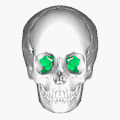
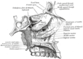
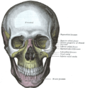
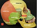
The sphenoid bone [note 1] is an unpaired bone of the neurocranium. It is situated in the middle of the skull towards the front, in front of the basilar part of the occipital bone. The sphenoid bone is one of the seven bones that articulate to form the orbit. Its shape somewhat resembles that of a butterfly or bat with its wings extended.

Contents

Structure

Seven bones articulate to form the orbit. The sphenoid bone is in red (directly in the middle of the orbital cavity) Orbital bones.png
Seven bones articulate to form the orbit. The sphenoid bone is in red (directly in the middle of the orbital cavity)
Wedge-shaped sphenoid bone seen laterally Gray164.png
Wedge-shaped sphenoid bone seen laterally

It is divided into the following parts:

Two sphenoidal conchae are situated at the anterior and inferior part of the body.

Intrinsic ligaments of the sphenoid

The more important of these are:

These ligaments occasionally ossify, though the incidence of ligamentous ossification (both partial and complete) varies according to the ligament type, with the interclinoid ligament being most commonly identified as having ossified and the pterygoalar ligament least commonly identified. [6]

Features

Articulations

The sphenoid articulates with the frontal, parietal, ethmoid, temporal, zygomatic, palatine, vomer, and occipital bones and helps to connect the neurocranium to the facial skeleton.

Body of sphenoid

Superior or cerebral surface

Articulates with ethmoid bone anteriorly and basilar part of occipital bone posteriorly. It shows:

  1. Jugum sphenoidale
  2. Sulcus chiasmaticus
  3. Tuberculum sellae
  4. Sella turcica
  5. Dorsum sellae
  6. Clivus [4]

Inferior surface

  1. Rostrum of sphenoid
  2. Sphenoidal conchae
  3. Vaginal processes of medial pterygoid plate

Anterior surface

Sphenoidal crest articulates with the perpendicular plate of ethmoid leading to formation of a part of the septum of nose.

Posterior surface

Basilar part of occipital bone

Lateral surface

Carotid sulcus lodging cavernous sinus and internal carotid artery

Sphenoidal sinuses

Sphenoidal sinuses are asymmetrical air sinuses in the body of the sphenoid, closed by sphenoidal conchae.

Greater wings

Superior or cerebral surface

This forms the floor of the middle cranial fossa. It presents (starting from the front):

Lateral surface

This is divided into (by infratemporal crest):

Foramen pierce it:

Orbital surface

This forms the posterior wall of the orbit [4]

Lesser wings

These are two triangular wings projecting laterally from anterosuperior part of the body. Each consists of:

Development

Figure 4 : Sphenoid bone at birth, posterior aspect. Gray148.png
Figure 4 : Sphenoid bone at birth, posterior aspect.

Until the seventh or eighth month of fetal development, the body of the sphenoid consists of two parts: one in front of the tuberculum sellæ, the presphenoid, with which the small wings are continuous; the other, consisting of the sella turcica and dorsum sellae, the postsphenoid, with which are associated the great wings, and pterygoid processes.

The greater part of the bone is ossified in cartilage. There are fourteen centers in all, six for the presphenoid and eight for the postsphenoid.

Presphenoid

By about the ninth week of fetal development an ossific center appears for each of the small wings (orbito-sphenoids) just lateral to the optic foramen; this is followed by the appearance of two nuclei in the presphenoid part of the body.

The sphenoidal conchae are each developed from a center that makes its appearance about the fifth month; at birth they consist of small triangular laminae, and it is not until the third year that they become hollowed out and coneshaped; about the fourth year they fuse with the labyrinths of the ethmoid bone, and between the ninth and twelfth years they unite with the sphenoid bone.

Postsphenoid

The first ossific nuclei are those for the great wings (alisphenoids). One makes its appearance in each wing between the foramen rotundum and foramen ovale about the eighth week. The orbital plate and that part of the sphenoid, which is found in the temporal fossa, as well as the lateral pterygoid plate, are ossified in membrane (Fawcett).

Soon after, the centers for the postsphenoid part of the body appear, one on either side of the sella turcica, and become blended together about the middle of fetal life.

Each medial pterygoid plate (except its hamulus) is ossified in membrane, and its center probably appears about the ninth or tenth week; the hamulus becomes chondrified during the third month, and almost at once ossifies (Fawcett).

The medial joins the lateral pterygoid plate about the sixth month.

About the fourth month, a center appears for each lingula and speedily joins the rest of the bone.

The presphenoid is united to the postsphenoid about the eighth month, and at birth the sphenoid is in three pieces [Fig. 4]: a central, consisting of the body and small wings, and two lateral, each comprising a great wing and pterygoid process.

In the first year after birth the great wings and body unite, and the small wings extend inward above the anterior part of the body, and, meeting with each other in the middle line, form an elevated smooth surface, termed the jugum sphenoidale.

By the twenty-fifth year the sphenoid and occipital are completely fused.

Between the pre- and postsphenoid there are occasionally seen the remains of a canal, the canalis cranio-pharyngeus, through which, in early fetal life, the hypophyseal diverticulum of the buccal ectoderm is transmitted.

The sphenoidal sinuses are present as minute cavities at the time of birth (Onodi), but do not attain their full size until after puberty.

Function

This bone assists with the formation of the base and the sides of the skull, and the floors and walls of the orbits. It is the site of attachment for most of the muscles of mastication. Many foramina and fissures are located in the sphenoid that carry nerves and blood vessels of the head and neck, such as the superior orbital fissure (with ophthalmic nerve), foramen rotundum (with maxillary nerve) and foramen ovale (with mandibular nerve). [7]

Other animals

The sphenoid bone of humans is homologous with a number of bones that are often separate in other animals, and have a somewhat complex arrangement.

In the early lobe-finned fishes and tetrapods, the pterygoid bones were flat, wing-like bones forming the major part of the roof of the mouth. Above the pterygoids were the epipterygoid bones, which formed part of a flexible joint between the braincase and the palatal region, as well as extending a vertical bar of bone towards the roof of the skull. Between the pterygoids lay an elongated, narrow parasphenoid bone, which also spread over some of the lower surface of the braincase, and connected, at its forward end, with a sphenethmoid bone helping to protect the olfactory nerves. Finally, the basisphenoid bone formed part of the floor of the braincase and lay immediately above the parasphenoid. [8]

Aside from the loss of the flexible joint at the rear of the palate, this primitive pattern is broadly retained in reptiles, albeit with some individual modifications. In birds, the epipterygoids are absent and the pterygoids considerably reduced. Living amphibians have a relatively simplified skull in this region; a broad parasphenoid forms the floor of the braincase, the pterygoids are relatively small, and all other related bones except the sphenethmoid are absent. [8]

In mammals, these various bones are often (though not always) fused into a single structure; the sphenoid. The basisphenoid forms the posterior part of the base, while the pterygoid processes represent the pterygoid bones. The epipterygoids have extended into the wall of the cranium; they are referred to as alisphenoids when separate in mammals, and form the greater wings of the sphenoid when fused into a larger structure. The sphenethmoid bone forms as three bones: the lesser wings and the anterior part of the base. These two parts of the sphenethmoid may be distinguished as orbitosphenoids and presphenoid, respectively, although there is often some degree of fusion. Only the parasphenoid appears to be entirely absent in mammals. [8]

In the dog the sphenoid is represented by 8 bones: basisphenoid, alisphenoids, presphenoid, orbitosphenoids, pterygoids. These bones remain separate and are the:

Additional images

See also

Notes

  1. According to most dictionaries, the word sphenoid ( /ˈsfnɔɪd/ [1] [2] ) derives from Greek sphenoeides, "wedgelike". Thieme Atlas of Anatomy [3] disagrees and says that the sphenoid bone was originally called os sphecoidale, meaning "bone resembling a wasp", and that the word was later written 'sphenoidale' by a transcription error. An anterior view of the bone resembles more the body of a wasp or a bat [4] with wings than a wedge.

Related Research Articles

<span class="mw-page-title-main">Maxilla</span> Upper jaw bone

In vertebrates, the maxilla is the upper fixed bone of the jaw formed from the fusion of two maxillary bones. In humans, the upper jaw includes the hard palate in the front of the mouth. The two maxillary bones are fused at the intermaxillary suture, forming the anterior nasal spine. This is similar to the mandible, which is also a fusion of two mandibular bones at the mandibular symphysis. The mandible is the movable part of the jaw.

<span class="mw-page-title-main">Zygomatic bone</span> Facial bone

In the human skull, the zygomatic bone, also called cheekbone or malar bone, is a paired irregular bone, situated at the upper and lateral part of the face and forms part of the lateral wall and floor of the orbit, of the temporal fossa and the infratemporal fossa. It presents a malar and a temporal surface; four processes, and four borders.

<span class="mw-page-title-main">Ethmoid bone</span> Bone of the facial skeletonl

The ethmoid bone is an unpaired bone in the skull that separates the nasal cavity from the brain. It is located at the roof of the nose, between the two orbits. The cubical bone is lightweight due to a spongy construction. The ethmoid bone is one of the bones that make up the orbit of the eye.

<span class="mw-page-title-main">Palatine bone</span> Bone of the facial skeleton

In anatomy, the palatine bones are two irregular bones of the facial skeleton in many animal species, located above the uvula in the throat. Together with the maxilla, they comprise the hard palate.

<span class="mw-page-title-main">Sella turcica</span> Saddle-shaped depression in the sphenoid bone of the skull

The sella turcica is a saddle-shaped depression in the body of the sphenoid bone of the human skull and of the skulls of other hominids including chimpanzees, gorillas and orangutans. It serves as a cephalometric landmark. The pituitary gland or hypophysis is located within the most inferior aspect of the sella turcica, the hypophyseal fossa.

<span class="mw-page-title-main">Posterior cranial fossa</span> Area of the cranium containing the brainstem and cerebellum

The posterior cranial fossa is the part of the cranial cavity located between the foramen magnum, and tentorium cerebelli. It is formed by the sphenoid bones, temporal bones, and occipital bone. It lodges the cerebellum, and parts of the brainstem.

<span class="mw-page-title-main">Cavernous sinus</span> Sinus in the human head

The cavernous sinus within the human head is one of the dural venous sinuses creating a cavity called the lateral sellar compartment bordered by the temporal bone of the skull and the sphenoid bone, lateral to the sella turcica.

<span class="mw-page-title-main">Sphenoid sinus</span> One of the four paired paranasal sinuses

The sphenoid sinus is a paired paranasal sinus occurring within the body of the sphenoid bone. It represents one pair of the four paired paranasal sinuses. The pair of sphenoid sinuses are separated in the middle by a septum of sphenoid sinuses. Each sphenoid sinus communicates with the nasal cavity via the opening of sphenoidal sinus. The two sphenoid sinuses vary in size and shape, and are usually asymmetrical.

<span class="mw-page-title-main">Sphenoidal conchae</span>

The sphenoidal conchae are two thin, curved plates, situated at the anterior and lower part of the body of the sphenoid. An aperture of variable size exists in the anterior wall of each, and through this the sphenoidal sinus opens into the nasal cavity.

<span class="mw-page-title-main">Greater wing of sphenoid bone</span> Large part of the skull front behind the eye socket

The greater wing of the sphenoid bone, or alisphenoid, is a bony process of the sphenoid bone, positioned in the skull behind each eye. There is one on each side, extending from the side of the body of the sphenoid and curving upward, laterally, and backward.

<span class="mw-page-title-main">Lesser wing of sphenoid bone</span>

The lesser wings of the sphenoid or orbito-sphenoids are two thin triangular plates, which arise from the upper and anterior parts of the body, and, projecting lateralward, end in sharp points [Fig. 1].

<span class="mw-page-title-main">Middle cranial fossa</span>

The middle cranial fossa is formed by the sphenoid bones, and the temporal bones. It lodges the temporal lobes, and the pituitary gland. It is deeper than the anterior cranial fossa, is narrow medially and widens laterally to the sides of the skull. It is separated from the posterior cranial fossa by the clivus and the petrous crest.

<span class="mw-page-title-main">Dorsum sellae</span>

The dorsum sellae is part of the sphenoid bone in the skull. Together with the basilar part of the occipital bone it forms the clivus.

<span class="mw-page-title-main">Posterior clinoid processes</span>

The posterior clinoid processes are the tubercles of the sphenoid bone situated at the superior angles of the dorsum sellæ which represents the posterior boundary of the sella turcica. They vary considerably size and form. The posterior clinoid processes deepen the sella turcica, and give attachment to the tentorium cerebelli, and the dura forming the floor of the hypophyseal fossa.

<span class="mw-page-title-main">Sphenoidal process of palatine bone</span> Thin, compressed plate

The sphenoidal process of palatine bone is a thin, superomedially directed plate of bone. It is smaller and more inferior compared to the orbital process of palatine bone.

<span class="mw-page-title-main">Perpendicular plate of palatine bone</span>

The perpendicular plate of palatine bone is the vertical part of the palatine bone, and is thin, of an oblong form, and presents two surfaces and four borders.

<span class="mw-page-title-main">Body of sphenoid bone</span>

The body of the sphenoid bone, more or less cubical in shape, is hollowed out in its interior to form two large cavities, the sphenoidal sinuses, which are separated from each other by a septum.

<span class="mw-page-title-main">Base of skull</span> Inferior area of the skull, composed of the endocranium and lower parts of the skull roof

The base of skull, also known as the cranial base or the cranial floor, is the most inferior area of the skull. It is composed of the endocranium and the lower parts of the calvaria.

<span class="mw-page-title-main">Outline of human anatomy</span> Overview of and topical guide to human anatomy

The following outline is provided as an overview of and topical guide to human anatomy:

<span class="mw-page-title-main">Neurocranium</span> Part of the skull around the brain

In human anatomy, the neurocranium, also known as the braincase, brainpan, or brain-pan, is the upper and back part of the skull, which forms a protective case around the brain. In the human skull, the neurocranium includes the calvaria or skullcap. The remainder of the skull is the facial skeleton.

References

  1. OED 2nd edition, 1989.
  2. Entry "sphenoid" in Merriam-Webster Online Dictionary .
  3. Thieme Atlas of Anatomy, Head and Neuroanatomy 2011
  4. 1 2 3 4 5 6 7 8 Chaurasia (31 January 2013). Human Anatomy Volume Three. CBS Publishers & Distributors. pp. 43–45. ISBN   978-81-239-2332-1.
  5. Jacob (2008). Human Anatomy. Elsevier. p. 211. ISBN   978-0-443-10373-5.
  6. Touska, P., Hasso, S., Oztek, A. et al. Skull base ligamentous mineralisation: evaluation using computed tomography and a review of the clinical relevance. Insights Imaging 10, 55 (2019). https://doi.org/10.1186/s13244-019-0740-8
  7. Fehrenbach; Herring (2012). Illustrated Anatomy of the Head and Neck. Elsevier. p. 52. ISBN   978-1-4377-2419-6.
  8. 1 2 3 Romer, Alfred Sherwood; Parsons, Thomas S. (1977). The Vertebrate Body. Philadelphia, PA: Holt-Saunders International. pp. 220–244. ISBN   0-03-910284-X.

PD-icon.svgThis article incorporates text in the public domain from page 147 of the 20th edition of Gray's Anatomy (1918)