Pterygoid processes of the sphenoid

Last updated
Pterygoid processes of the sphenoid
Gray145.png
Sphenoid bone, upper surface.
Gray146.png
Sphenoid bone, anterior and inferior surfaces.
Details
Identifiers
Latin processus pterygoideus ossis sphenoidalis
TA98 A02.1.05.042
TA2 628
FMA 54682
Anatomical terms of bone

The pterygoid processes of the sphenoid (from Greek pteryx, pterygos, "wing"), one on either side, descend perpendicularly from the regions where the body and the greater wings of the sphenoid bone unite.

Contents

Each process consists of a medial pterygoid plate and a lateral pterygoid plate, the latter of which serve as the origins of the medial and lateral pterygoid muscles. The medial pterygoid, along with the masseter allows the jaw to move in a vertical direction as it contracts and relaxes. The lateral pterygoid allows the jaw to move in a horizontal direction during mastication (chewing). Fracture of either plate are used in clinical medicine to distinguish the Le Fort fracture classification for high impact injuries to the sphenoid and maxillary bones.

The superior portion of the pterygoid processes are fused anteriorly; a vertical groove, the pterygopalatine fossa, descends on the front of the line of fusion. The plates are separated below by an angular cleft, the pterygoid notch, the margins of which are rough for articulation with the pyramidal process of the palatine bone.

The two plates diverge behind and enclose between them a V-shaped fossa, the pterygoid fossa, which contains the medial pterygoid muscle and the tensor veli palatini.

Above this fossa is a small, oval, shallow depression, the scaphoid fossa, which gives origin to the tensor veli palatini.

The anterior surface of the pterygoid process is broad and triangular near its root, where it forms the posterior wall of the pterygopalatine fossa and presents the anterior orifice of the pterygoid canal.

In many mammals it remains as a separate bone called the pterygoid bone.

Its name is Greek for "resembling a fin or wing", from its shape.

Medial pterygoid plate

The medial pterygoid plate (or medial pterygoid lamina) of the sphenoid bone is a horse-shoe shaped process that arises from its underside.

It is narrower and longer than the lateral pterygoid plate and curves lateralward at its lower extremity into a hook-like process, the pterygoid hamulus, around which the tendon of the tensor veli palatini glides.

The lateral surface of this plate forms part of the pterygoid fossa, the medial surface constitutes the lateral boundary of the choana or posterior aperture of the corresponding nasal cavity.

Superiorly the medial plate is prolonged on to the under surface of the body as a thin lamina, named the vaginal process, which articulates in front with the sphenoidal process of the palatine and behind this with the ala (wing) of the vomer.

The angular prominence between the posterior margin of the vaginal process and the medial border of the scaphoid fossa is named the pterygoid tubercle, and immediately above this is the posterior opening of the pterygoid canal.

On the under surface of the vaginal process is a furrow, which is converted into a canal by the sphenoidal process of the palatine bone, for the transmission of the pharyngeal branch of the internal maxillary artery and the pharyngeal nerve from the sphenopalatine ganglion.

The pharyngeal aponeurosis is attached to the entire length of the posterior edge of the medial plate, and the constrictor pharyngis superior takes origin from its lower third.

Projecting backward from near the middle of the posterior edge of this plate is an angular process, the processus tubarius, which supports the pharyngeal end of the Eustachian tube.

The anterior margin of the plate articulates with the posterior border of the vertical part of the palatine bone.

In many animals it is a separate bone called the pterygoid bone.

Lateral pterygoid plate

Lateral pterygoid plate
Gray192.png
Medial wall of left orbit. (Lateral pterygoid plate labeled at bottom right.)
Details
Identifiers
Latin lamina lateralis processus pterygoidei
TA98 A02.1.05.042
TA2 628
FMA 54682
Anatomical terms of bone

The lateral pterygoid plate of the sphenoid (or lateral lamina of pterygoid process) is broad, thin, and everted and forms the lateral part of a horseshoe like process that extends from the inferior aspect of the sphenoid bone, and serves as the origin of the lateral pterygoid muscle, which functions in allowing the mandible to move in a lateral and medial direction, or from side-to-side.

Its lateral surface forms part of the medial wall of the infratemporal fossa, and gives attachment to the lateral pterygoid muscle; its medial surface forms part of the pterygoid fossa, and gives attachment to the medial pterygoid muscle. Posterior edge is sharp, and often has sharp projection - pterygospinous process (Civinini process).

Related Research Articles

Articles related to anatomy include:

<span class="mw-page-title-main">Sphenoid bone</span> Bone at the front of the skull

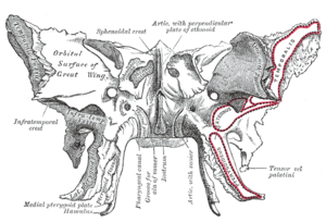
The sphenoid bone is an unpaired bone of the neurocranium. It is situated in the middle of the skull towards the front, in front of the basilar part of the occipital bone. The sphenoid bone is one of the seven bones that articulate to form the orbit. Its shape somewhat resembles that of a butterfly or bat with its wings extended.

<span class="mw-page-title-main">Palatine bone</span> Bone of the facial skeleton

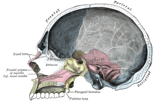
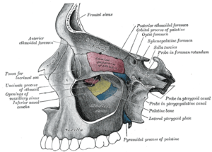
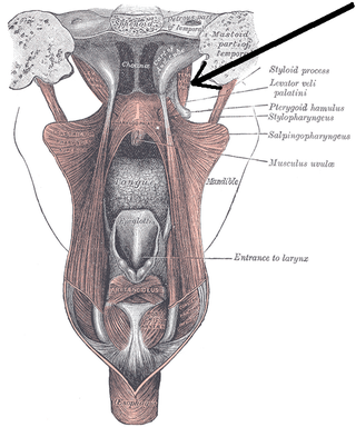
In anatomy, the palatine bones are two irregular bones of the facial skeleton in many animal species, located above the uvula in the throat. Together with the maxillae, they comprise the hard palate.

<span class="mw-page-title-main">Orbit (anatomy)</span> Cavity or socket of the skull in which the eye and its appendages are situated

In anatomy, the orbit is the cavity or socket/hole of the skull in which the eye and its appendages are situated. "Orbit" can refer to the bony socket, or it can also be used to imply the contents. In the adult human, the volume of the orbit is 30 millilitres, of which the eye occupies 6.5 ml. The orbital contents comprise the eye, the orbital and retrobulbar fascia, extraocular muscles, cranial nerves II, III, IV, V, and VI, blood vessels, fat, the lacrimal gland with its sac and duct, the eyelids, medial and lateral palpebral ligaments, cheek ligaments, the suspensory ligament, septum, ciliary ganglion and short ciliary nerves.

<span class="mw-page-title-main">Medial pterygoid muscle</span> Muscle involved in chewing

The medial pterygoid muscle, is a thick, quadrilateral muscle of the face. It is supplied by the mandibular branch of the trigeminal nerve (V). It is important in mastication (chewing).

<span class="mw-page-title-main">Levator veli palatini</span> Muscle of the soft palate

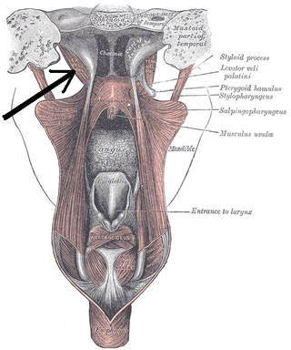
The levator veli palatini is a muscle of the soft palate and pharynx. It is innervated by the vagus nerve via its pharyngeal plexus. During swallowing, it contracts, elevating the soft palate to help prevent food from entering the nasopharynx.

<span class="mw-page-title-main">Tensor veli palatini muscle</span> Muscle of the soft palate

The tensor veli palatini muscle is a thin, triangular muscle of the head that tenses the soft palate and opens the Eustachian tube to equalise pressure in the middle ear.

<span class="mw-page-title-main">Pterygopalatine fossa</span> Fossa in the skull

In human anatomy, the pterygopalatine fossa is a fossa in the skull. A human skull contains two pterygopalatine fossae—one on the left side, and another on the right side. Each fossa is a cone-shaped paired depression deep to the infratemporal fossa and posterior to the maxilla on each side of the skull, located between the pterygoid process and the maxillary tuberosity close to the apex of the orbit. It is the indented area medial to the pterygomaxillary fissure leading into the sphenopalatine foramen. It communicates with the nasal and oral cavities, infratemporal fossa, orbit, pharynx, and middle cranial fossa through eight foramina.

<span class="mw-page-title-main">Sphenoidal conchae</span>

The sphenoidal conchae are two thin, curved plates, situated at the anterior and lower part of the body of the sphenoid. An aperture of variable size exists in the anterior wall of each, and through this the sphenoidal sinus opens into the nasal cavity.

<span class="mw-page-title-main">Greater wing of sphenoid bone</span> Large part of the skull front behind the eye socket

The greater wing of the sphenoid bone, or alisphenoid, is a bony process of the sphenoid bone; there is one on each side, extending from the side of the body of the sphenoid and curving upward, laterally, and backward.

<span class="mw-page-title-main">Ethmoidal labyrinth</span>

The ethmoidal labyrinth or lateral mass of the ethmoid bone consists of a number of thin-walled cellular cavities, the ethmoid air cells, arranged in three groups, anterior, middle, and posterior, and interposed between two vertical plates of bone; the lateral plate forms part of the orbit, the medial plate forms part of the nasal cavity. In the disarticulated bone many of these cells are opened into, but when the bones are articulated, they are closed in at every part, except where they open into the nasal cavity.

<span class="mw-page-title-main">Maxillary artery</span> Artery supplying face structures in humans

The maxillary artery supplies deep structures of the face. It branches from the external carotid artery just deep to the neck of the mandible.

<span class="mw-page-title-main">Middle cranial fossa</span>

The middle cranial fossa is formed by the sphenoid bones, and the temporal bones. It lodges the temporal lobes, and the pituitary gland. It is deeper than the anterior cranial fossa, is narrow medially and widens laterally to the sides of the skull. It is separated from the posterior cranial fossa by the clivus and the petrous crest.

<span class="mw-page-title-main">Infratemporal fossa</span> Cavity that is part of the skull

The infratemporal fossa is an irregularly shaped cavity that is a part of the skull. It is situated below and medial to the zygomatic arch. It is not fully enclosed by bone in all directions. It contains superficial muscles, including the lower part of the temporalis muscle, the lateral pterygoid muscle, and the medial pterygoid muscle. It also contains important blood vessels such as the middle meningeal artery, the pterygoid plexus, and the retromandibular vein, and nerves such as the mandibular nerve (CN V3) and its branches.

<span class="mw-page-title-main">Palatovaginal canal</span>

The palatovaginal canal is a small canal formed between the sphenoidal process of palatine bone, and vaginal process of sphenoid bone. It connects the pterygopalatine fossa and and nasal cavity. It transmits the pharyngeal nerve, and the pharyngeal branch of maxillary artery.

<span class="mw-page-title-main">Sphenoidal process of palatine bone</span> Thin, compressed plate

The sphenoidal process of palatine bone is a thin, superomedially directed plate of bone. It is smaller and more inferior compared to the orbital process of palatine bone.

<span class="mw-page-title-main">Perpendicular plate of palatine bone</span>

The perpendicular plate of palatine bone is the vertical part of the palatine bone, and is thin, of an oblong form, and presents two surfaces and four borders.

<span class="mw-page-title-main">Pterygoid fossa</span>

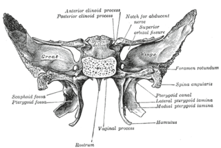
The pterygoid fossa is an anatomical term for the fossa formed by the divergence of the lateral pterygoid plate and the medial pterygoid plate of the sphenoid bone.

<span class="mw-page-title-main">Body of sphenoid bone</span>

The body of the sphenoid bone, more or less cubical in shape, is hollowed out in its interior to form two large cavities, the sphenoidal sinuses, which are separated from each other by a septum.

<span class="mw-page-title-main">Outline of human anatomy</span> Overview of and topical guide to human anatomy

The following outline is provided as an overview of and topical guide to human anatomy:

References

PD-icon.svgThis article incorporates text in the public domain from page 151 of the 20th edition of Gray's Anatomy (1918)