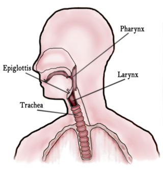
In vertebrate anatomy, the throat is the front part of the neck, internally positioned in front of the vertebrae. It contains the pharynx and larynx. An important section of it is the epiglottis, separating the esophagus from the trachea (windpipe), preventing food and drinks being inhaled into the lungs. The throat contains various blood vessels, pharyngeal muscles, the nasopharyngeal tonsil, the tonsils, the palatine uvula, the trachea, the esophagus, and the vocal cords. Mammal throats consist of two bones, the hyoid bone and the clavicle. The "throat" is sometimes thought to be synonymous for the fauces.

A joint or articulation is the connection made between bones, ossicles, or other hard structures in the body which link an animal's skeletal system into a functional whole. They are constructed to allow for different degrees and types of movement. Some joints, such as the knee, elbow, and shoulder, are self-lubricating, almost frictionless, and are able to withstand compression and maintain heavy loads while still executing smooth and precise movements. Other joints such as sutures between the bones of the skull permit very little movement in order to protect the brain and the sense organs. The connection between a tooth and the jawbone is also called a joint, and is described as a fibrous joint known as a gomphosis. Joints are classified both structurally and functionally.

The brachioradialis is a muscle of the forearm that flexes the forearm at the elbow. It is also capable of both pronation and supination, depending on the position of the forearm. It is attached to the distal styloid process of the radius by way of the brachioradialis tendon, and to the lateral supracondylar ridge of the humerus.

A pulmonary artery is an artery in the pulmonary circulation that carries deoxygenated blood from the right side of the heart to the lungs. The largest pulmonary artery is the main pulmonary artery or pulmonary trunk from the heart, and the smallest ones are the arterioles, which lead to the capillaries that surround the pulmonary alveoli.

Morphology is a branch of biology dealing with the study of the form and structure of organisms and their specific structural features.

The internal pudendal artery is one of the three pudendal arteries. It branches off the internal iliac artery, and provides blood to the external genitalia.

The mediastinum is the central compartment of the thoracic cavity. Surrounded by loose connective tissue, it is an undelineated region that contains a group of structures within the thorax, namely the heart and its vessels, the esophagus, the trachea, the phrenic and cardiac nerves, the thoracic duct, the thymus and the lymph nodes of the central chest.
Grey's Anatomy is an American medical drama television series focusing on the personal and professional lives of surgical interns, residents, and attendings at the fictional Seattle Grace Hospital, later named the Grey Sloan Memorial Hospital. The series premiered on March 27, 2005, on ABC as a mid-season replacement. The show's title is an allusion to Gray's Anatomy, a classic human anatomy textbook. Writer Shonda Rhimes developed the pilot and served as showrunner, head writer, and executive producer until stepping down in 2015. Set in Seattle, Washington, the series is filmed primarily in Los Angeles, California, and Vancouver, British Columbia.

In human anatomy, the groin, also known as the inguinal region or iliac region, is the junctional area between the torso and the thigh. The groin is at the front of the body on either side of the pubic tubercle, where the lower part of the abdominal wall meets the thigh. A fold or crease is formed at this junction known as the inguinal groove, or crease. This is also the area of the medial compartment of the thigh that contains attachments of the adductor muscles of the hip or the groin muscles. The groin is the common site for a hernia.

The celiacartery, also known as the celiac trunk or truncus coeliacus, is the first major branch of the abdominal aorta. It is about 1.25 cm in length. Branching from the aorta at thoracic vertebra 12 (T12) in humans, it is one of three anterior/ midline branches of the abdominal aorta.

The olecranon, is a large, thick, curved bony process on the proximal, posterior end of the ulna. It forms the protruding part of the elbow and is opposite to the cubital fossa or elbow pit. The olecranon serves as a lever for the extensor muscles that straighten the elbow joint.

The temple, also known as the pterion, is a latch where four skull bones fuse: the frontal, parietal, temporal, and sphenoid. It is located on the side of the head behind the eye between the forehead and the ear. The temporal muscle covers this area and is used during mastication.

The semispinalis muscles are a group of three muscles belonging to the transversospinales. These are the semispinalis capitis, the semispinalis cervicis and the semispinalis thoracis.
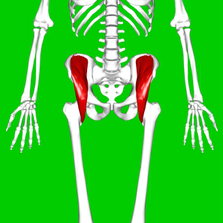
The iliacus is a flat, triangular muscle which fills the iliac fossa. It forms the lateral portion of iliopsoas, providing flexion of the thigh and lower limb at the acetabulofemoral joint.

The stylomastoid foramen is a foramen between the styloid and mastoid processes of the temporal bone of the skull. It is the termination of the facial canal, and transmits the facial nerve, and stylomastoid artery. Facial nerve inflammation in the stylomastoid foramen may cause Bell's palsy.

The vaginal artery is an artery in females that supplies blood to the vagina and the base of the bladder.

The inferior rectal artery is an artery that supplies blood to the lower third of the anal canal below the pectinate line.
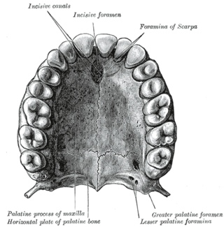
In the human mouth, the incisive foramen is the opening of the incisive canals on the hard palate immediately behind the incisor teeth. It gives passage to blood vessels and nerves. The incisive foramen is situated within the incisive fossa of the maxilla.

The broad ligament of the uterus is the wide fold of peritoneum that connects the sides of the uterus to the walls and floor of the pelvis.

Each vertebra is an irregular bone with a complex structure composed of bone and some hyaline cartilage, that make up the vertebral column or spine, of vertebrates. The proportions of the vertebrae differ according to their spinal segment and the particular species.

