Anatomical terms of bone

Last updated

Many anatomical terms descriptive of bone are defined in anatomical terminology, and are often derived from Greek and Latin. Bone in the human body is categorized into long bone, short bone, flat bone, irregular bone and sesamoid bone.

Contents

Types of bone

Different types of bone 601 Bone Classification.jpg
Different types of bone

Long bones

A long bone is one that is cylindrical in shape, being longer than it is wide. However, the term describes the shape of a bone, not its size, which is relative. Long bones are found in the arms (humerus, ulna, radius) and legs (femur, tibia, fibula), as well as in the fingers (metacarpals, phalanges) and toes (metatarsals, phalanges). Long bones function as levers; they move when muscles contract. [1]

Short bones

A short bone is one that is cube-like in shape, being approximately equal in length, width, and thickness. The only short bones in the human skeleton are in the carpals of the wrists and the tarsals of the ankles. Short bones provide stability and support as well as some limited motion. [1]

Flat bones

The term “flat bone” is something of a misnomer because, although a flat bone is typically thin, it is also often curved. Examples include the cranial (skull) bones, the scapulae (shoulder blades), the sternum (breastbone), and the ribs. Flat bones serve as points of attachment for muscles and often protect internal organs. Flat bones do not have a medullary cavity because they are thin. [1]

Irregular bones

An irregular bone is one that does not have an easily classified shape and defies description. These bones tend to have more complex shapes, like the vertebrae that support the spinal cord and protect it from compressive forces. Many facial bones, particularly the ones containing sinuses, are classified as irregular bones. [1]

Sesamoid bones

A sesamoid bone is a small, round bone that, as the name suggests, is shaped like a sesame seed. These bones form in tendons (the sheaths of tissue that connect bones to muscles) where a great deal of pressure is generated in a joint. The sesamoid bones protect tendons by helping them overcome compressive forces. Sesamoid bones vary in number and placement from person to person but are typically found in tendons associated with the feet, hands, and knees. The only type of sesamoid bone that is common to everybody is the kneecap (patella, pl. patellae) which is also the largest of the sesamoid bones. [1]

Protrusions

Rounded

Different types of bone markings and features. 602 Bone Markings.jpg
Different types of bone markings and features.

A condyle is the round prominence at the end of a bone, most often part of a joint – an articulation with another bone. [2] The epicondyle refers to a projection near a condyle, particularly the medial epicondyle of the humerus. [3] These terms derive from Greek. [4] [lower-alpha 1]

An eminence refers to a relatively small projection or bump, particularly of bone, such as the medial eminence. [5]

A process refers to a relatively large projection or prominent bump, [6] as does a promontory such as the sacral promontory. [7]

Both tubercle and tuberosity refer to a projection or bump with a roughened surface, with a "tubercle" generally smaller than a "tuberosity". These terms are derived from tuber (Latin : swelling). [8] , as is also protuberance , which occasionally is synonymous with "tuberosity".

A ramus (Latin : branch) refers to an extension of bone, [9] such as the ramus of the mandible in the jaw or superior pubic ramus. Ramus may also be used to refer to nerves, such as the ramus communicans.

A facet refers to a small, flattened articular surface.[ citation needed ]

Pointed

A line refers to a long, thin projection, often with a rough surface.

Ridge and crest refer to a long, narrow line. [10] Unlike many words used to describe anatomical terms, the word ridge is derived from Old English. [11] [lower-alpha 2]

A spine, as well as referring to the spinal cord, may be used to describe a relatively long, thin projection or bump.

Special

These terms are used to describe bony protuberances in specific parts of the body.

The Malleolus (Latin : "small hammer") is the bony prominence on each side of the ankle. [12] These are known as the medial and lateral malleolus. Each leg is supported by two bones, the tibia on the inner side (medial) of the leg and the fibula on the outer side (lateral) of the leg. The medial malleolus is the prominence on the inner side of the ankle, formed by the lower end of the tibia. The lateral malleolus is the prominence on the outer side of the ankle, formed by the lower end of the fibula.

The trochanters are parts of the femur, to which muscles attach. [13] It may refer to the greater, lesser, or third trochanter

Cavities

Openings

The following terms are used to describe cavities that connect to other areas:

A foramen is any opening, particularly referring to those in bone. [14] Foramina inside the body of humans and other animals typically allow muscles, nerves, arteries, veins, or other structures to connect one part of the body with another. An example is the foramen magnum in occipital bone.

A canal is a long, tunnel-like foramen, usually a passage for notable nerves or blood vessels. An example is the auditory canal.

Blind-ended

The following terms are used to describe cavities that do not connect to other areas:

A fossa (from the Latin "fossa", ditch or trench) is a depression or hollow, usually in a bone, such as the hypophyseal fossa, the depression in the sphenoid bone. [15]

A meatus is a short canal that opens to another part of the body. [16] An example is the external auditory meatus.

A fovea (Latin : pit) is a small pit, usually on the head of a bone. An example of a fovea is the fovea capitis of the head of the femur.

Walls

The following terms are used to describe the walls of a cavity:

A labyrinth refers to the bony labyrinth and membranous labyrinth, components of the inner ear, due to their fine and complex structure. [17]

A sinus refers to a bony cavity, usually within the skull. [18]

Joints

A joint, or articulation is the region where adjacent bones contact each other, for example the elbow, shoulder, or costovertebral joint. Terms that refer to joints include:

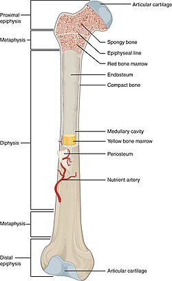
Features of long bones

Gross overview of the features of long bones in a fully grown adult. 603 Anatomy of Long Bone.jpg
Gross overview of the features of long bones in a fully grown adult.

Gross features

Bones are commonly described with the terms head, neck, shaft, body and base

The head of a bone usually refers to the distal end of the bone. The shaft refers to the elongated sections of long bone, and the neck the segment between the head and shaft (or body). The end of the long bone opposite to the head is known as the base.

Internal regions

The epiphyseal plate is the area in which bone growth occurs after birth through endochondral ossification. 623 Epiphyseal Plate-Line.jpg
The epiphyseal plate is the area in which bone growth occurs after birth through endochondral ossification.
Bone featureDefinitionEtymologic memory aid
apophysis Any of various processes or protuberances on a bone. apo- + physis , "outward from the growth part; outgrowth"
diaphysis The long, relatively straight main body of a long bone; region of primary ossification. Also known as the shaft. dia- + physis , "between the growth parts"
epiphysis The end regions of a long bone; regions of secondary ossification. epi- + physis , "on top of the growth part"
physis (epiphyseal plate)Also known as the growth plate. In a long bone it is a thin disc of hyaline cartilage that is positioned transversely between the epiphysis and metaphysis. In the long bones of humans, the epiphyseal plate disappears by twenty years of age. physis , "the growth part"
metaphysis The region of a long bone lying between the epiphysis and diaphysis. meta- + physis , "the transitional part (between shaft and end) leading to the growth part"

Internal and external

Inside of the head of femur, showing surface of the bone, red and yellow bone marrow. 619 Red and Yellow Bone Marrow.jpg
Inside of the head of femur, showing surface of the bone, red and yellow bone marrow.

The cortex of a bone is used to refer to its outer layers, and medulla used to refer to the inner surface of the bone. Red marrow, in which blood is formed is present in spongy bone as well as in the medullary cavity, while the fatty yellow marrow is present primarily in the medullary cavity. [ citation needed ]

See also

Notes

  1. Specifically, Latin: condylus, from "knuckle" (Greek: kondylos), with "epi" referring to "Upon". [4]
  2. Specifically, 'spine, crest' (Old English:hrycg) from Germanic related to the Dutch "rug" and German "Rücken" 'back'. [11]

Related Research Articles

<span class="mw-page-title-main">Human leg</span> Lower extremity or limb of the human body (foot, lower leg, thigh and hip)

The leg is the entire lower limb of the human body, including the foot, thigh or sometimes even the hip or buttock region. The major bones of the leg are the femur, tibia, and adjacent fibula. The thigh is between the hip and knee, while the calf (rear) and shin (front) are between the knee and foot.

<span class="mw-page-title-main">Femur</span> Thigh bone

The femur, or thigh bone is the only bone in the thigh. The thigh is the region of the lower limb between the hip and the knee. In many four-legged animals the femur is the upper bone of the hindleg.

<span class="mw-page-title-main">Temporomandibular joint</span> Joints connecting the jawbone to the skull

In anatomy, the temporomandibular joints (TMJ) are the two joints connecting the jawbone to the skull. It is a bilateral synovial articulation between the temporal bone of the skull above and the mandible below; it is from these bones that its name is derived. This joint is unique in that it is a bilateral joint that functions as one unit. Since the TMJ is connected to the mandible, the right and left joints must function together and therefore are not independent of each other.

<span class="mw-page-title-main">Scapula</span> Bone that connects the humerus (upper arm bone) with the clavicle (collar bone)

The scapula, also known as the shoulder blade, is the bone that connects the humerus with the clavicle. Like their connected bones, the scapulae are paired, with each scapula on either side of the body being roughly a mirror image of the other. The name derives from the Classical Latin word for trowel or small shovel, which it was thought to resemble.

Articles related to anatomy include:

<span class="mw-page-title-main">Humerus</span> Long bone of the upper arm

The humerus is a long bone in the arm that runs from the shoulder to the elbow. It connects the scapula and the two bones of the lower arm, the radius and ulna, and consists of three sections. The humeral upper extremity consists of a rounded head, a narrow neck, and two short processes. The body is cylindrical in its upper portion, and more prismatic below. The lower extremity consists of 2 epicondyles, 2 processes, and 3 fossae. As well as its true anatomical neck, the constriction below the greater and lesser tubercles of the humerus is referred to as its surgical neck due to its tendency to fracture, thus often becoming the focus of surgeons.

<span class="mw-page-title-main">Tibia</span> Leg bone in vertebrates

The tibia, also known as the shinbone or shankbone, is the larger, stronger, and anterior (frontal) of the two bones in the leg below the knee in vertebrates ; it connects the knee with the ankle. The tibia is found on the medial side of the leg next to the fibula and closer to the median plane. The tibia is connected to the fibula by the interosseous membrane of leg, forming a type of fibrous joint called a syndesmosis with very little movement. The tibia is named for the flute tibia. It is the second largest bone in the human body, after the femur. The leg bones are the strongest long bones as they support the rest of the body.

<span class="mw-page-title-main">Palatine bone</span> Bone of the facial skeleton

In anatomy, the palatine bones are two irregular bones of the facial skeleton in many animal species, located above the uvula in the throat. Together with the maxillae, they comprise the hard palate.

<span class="mw-page-title-main">Temporal bone</span> Sides and base of skull, connecting to the jaw and occipital bone

The temporal bones are situated at the sides and base of the skull, and lateral to the temporal lobes of the cerebral cortex.

<span class="mw-page-title-main">Obturator internus muscle</span> One of six small hip muscles in the lateral rotator group

The internal obturator muscle or obturator internus muscle originates on the medial surface of the obturator membrane, the ischium near the membrane, and the rim of the pubis.

<span class="mw-page-title-main">Adductor magnus muscle</span> Muscle in the thigh

The adductor magnus is a large triangular muscle, situated on the medial side of the thigh.

<span class="mw-page-title-main">Dog anatomy</span> Studies of the visible part of a canine

Dog anatomy comprises the anatomical studies of the visible parts of the body of a domestic dog. Details of structures vary tremendously from breed to breed, more than in any other animal species, wild or domesticated, as dogs are highly variable in height and weight. The smallest known adult dog was a Yorkshire Terrier that stood only 6.3 cm (2.5 in) at the shoulder, 9.5 cm (3.7 in) in length along the head and body, and weighed only 113 grams (4.0 oz). The heaviest dog was an English Mastiff named Zorba which weighed 314 pounds (142 kg). The tallest known adult dog is a Great Dane that stands 106.7 cm (42.0 in) at the shoulder.

<span class="mw-page-title-main">Ischium</span> Lower and back region of the hip bone

The ischium forms the lower and back region of the hip bone.

<span class="mw-page-title-main">Infratemporal fossa</span> Cavity that is part of the skull

The infratemporal fossa is an irregularly shaped cavity that is a part of the skull. It is situated below and medial to the zygomatic arch. It is not fully enclosed by bone in all directions. It contains superficial muscles, including the lower part of the temporalis muscle, the lateral pterygoid muscle, and the medial pterygoid muscle. It also contains important blood vessels such as the middle meningeal artery, the pterygoid plexus, and the retromandibular vein, and nerves such as the mandibular nerve (CN V3) and its branches.

<span class="mw-page-title-main">Outline of human anatomy</span> Overview of and topical guide to human anatomy

The following outline is provided as an overview of and topical guide to human anatomy:

<span class="mw-page-title-main">Hip bone</span> Bone of the pelvis

The hip bone is a large flat bone, constricted in the center and expanded above and below. In some vertebrates it is composed of three parts: the ilium, ischium, and the pubis.

<span class="mw-page-title-main">Mandible</span> Lower jaw bone

In jawed vertebrates, the mandible, lower jaw, or jawbone is a bone that makes up the lower – and typically more mobile – component of the mouth.

<span class="mw-page-title-main">Pelvis</span> Lower torso of the human body

The pelvis is the lower part of the trunk, between the abdomen and the thighs, together with its embedded skeleton.

This glossary explains technical terms commonly employed in the description of dinosaur body fossils. Besides dinosaur-specific terms, it covers terms with wider usage, when these are of central importance in the study of dinosaurs or when their discussion in the context of dinosaurs is beneficial. The glossary does not cover ichnological and bone histological terms, nor does it cover measurements.

In the skeleton of humans and other animals, a tubercle, tuberosity or apophysis is a protrusion or eminence that serves as an attachment for skeletal muscles. The muscles attach by tendons, where the enthesis is the connective tissue between the tendon and bone. A tuberosity is generally a larger tubercle.

References

This Wikipedia entry incorporates text from the freely licensed Connexions edition of Anatomy & Physiology text-book by OpenStax College

  1. 1 2 3 4 5 "Anatomy & Physiology". Openstax college at Connexions. Retrieved November 16, 2013.
  2. OED 1989, "Condyle".
  3. OED 1989, "Epicondyle".
  4. 1 2 OED 1989, "Condyle, Epicondyle, Epi-".
  5. OED 1989, "eminence".
  6. OED 1989, "process".
  7. OED 1989, "promontory".
  8. OED 1989, "Tuberous, Tubercle".
  9. OED 1989, "ramus".
  10. OED 1989, "Crest".
  11. 1 2 OED 1989, "Ridge".
  12. OED 1989, "Malleolus".
  13. OED 1989, "Trochanter".
  14. OED 1989, "Foramen".
  15. Venieratos D, Anagnostopoulou S, Garidou A., A new morphometric method for the sella turcica and the hypophyseal fossa and its clinical relevance. Folia Morphol (Warsz). 2005 Nov; 64(4): 240–47. PMID   16425149
  16. OED 1989, "meatus".
  17. OED 1989, "labyrinth".
  18. OED 1989, "sinus".
Books