Lesser trochanter

Last updated
Lesser trochanter
Gray341.png
Left hip-joint, opened by removing the floor of the acetabulum from within the pelvis.
Gray243.png
Upper extremity of right femur viewed from behind and above.
Details
Insertions Psoas major, Iliacus
Identifiers
Latin trochanter minor
TA98 A02.5.04.007
TA2 1366
FMA 32853
Anatomical terms of bone

In human anatomy, the lesser trochanter is a conical, posteromedial, bony projection from the shaft of the femur. it serves as the principal insertion site of the iliopsoas muscle. [1]

Contents

Structure

The lesser trochanter is a conical posteromedial projection of the shaft of the femur, projecting from the posteroinferior aspect of its junction with the femoral neck. [1]

The summit and anterior surface of the lesser trochanter are rough, whereas its posterior surface is smooth. [1]

From its apex three well-marked borders extend: [2]

Attachments

The summit of the lesser trochanter gives insertion to the tendon of the psoas major muscle and the iliacus muscle; [3] the lesser trochanter represents the principal attachment of the iliopsoas. [1]

Anatomical relations

The intertrochanteric crest (which demarcates the junction of the femoral shaft and neck posteriorly) extends between the lesser trochanter and the greater trochanter on the posterior surface of the femur. [1]

Clinical significance

The lesser trochanter can be involved in an avulsion fracture. [4]

Other animals

Paleontology

The position of the lesser trochanter close to the head of the femur is one of the defining characteristics of the Prozostrodontia, which is the clade of cynodonts including mammals and their closest non-mammaliform relatives. It was erected as a node-based taxon as the least inclusive clade containing Prozostrodon brasiliensis , Tritylodon langaevus , Pachygenelus monus , and Mus musculus (the house mouse). [5]

All living mammals have a lesser trochanter, whose size, shape, and position is distinctive to their species.

Additional images

See also

Related Research Articles

<span class="mw-page-title-main">Human leg</span> Lower extremity or limb of the human body (foot, lower leg, thigh and hip)

The leg is the entire lower limb of the human body, including the foot, thigh or sometimes even the hip or buttock region. The major bones of the leg are the femur, tibia, and adjacent fibula. The thigh is between the hip and knee, while the calf (rear) and shin (front) are between the knee and foot.

<span class="mw-page-title-main">Femur</span> Thigh bone

The femur, or thigh bone is the only bone in the thigh. The thigh is the region of the lower limb between the hip and the knee. In many four-legged animals the femur is the upper bone of the hindleg.

<span class="mw-page-title-main">Humerus</span> Long bone of the upper arm

The humerus is a long bone in the arm that runs from the shoulder to the elbow. It connects the scapula and the two bones of the lower arm, the radius and ulna, and consists of three sections. The humeral upper extremity consists of a rounded head, a narrow neck, and two short processes. The body is cylindrical in its upper portion, and more prismatic below. The lower extremity consists of 2 epicondyles, 2 processes, and 3 fossae. As well as its true anatomical neck, the constriction below the greater and lesser tubercles of the humerus is referred to as its surgical neck due to its tendency to fracture, thus often becoming the focus of surgeons.

<span class="mw-page-title-main">Greater trochanter</span> Quadrilateral part of the thigh bone

The greater trochanter of the femur is a large, irregular, quadrilateral eminence and a part of the skeletal system.

<span class="mw-page-title-main">Hip</span> Anatomical region between the torso and the legs, holding the buttocks and genital region

In vertebrate anatomy, the hip, or coxa(pl.: coxae) in medical terminology, refers to either an anatomical region or a joint on the outer (lateral) side of the pelvis.

<span class="mw-page-title-main">Psoas major muscle</span> Long fusiform muscle located in the lumbar region

The psoas major is a long fusiform muscle located in the lateral lumbar region between the vertebral column and the brim of the lesser pelvis. It joins the iliacus muscle to form the iliopsoas. In animals, this muscle is equivalent to the tenderloin.

<span class="mw-page-title-main">Iliacus muscle</span> Flat, triangular muscle which fills the iliac fossa

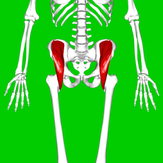
The iliacus is a flat, triangular muscle which fills the iliac fossa. It forms the lateral portion of iliopsoas, providing flexion of the thigh and lower limb at the acetabulofemoral joint.

<span class="mw-page-title-main">External obturator muscle</span> One of six small hip muscles in the lateral rotator group

The external obturator muscle or obturator externus muscle is a flat, triangular muscle, which covers the outer surface of the anterior wall of the pelvis.

<span class="mw-page-title-main">Quadratus femoris muscle</span> One of six small hip muscles in the lateral rotator group

The quadratus femoris is a flat, quadrilateral skeletal muscle. Located on the posterior side of the hip joint, it is a strong external rotator and adductor of the thigh, but also acts to stabilize the femoral head in the acetabulum. The quadratus femoris is used in Meyer's muscle pedicle grafting to prevent avascular necrosis of femur head.

<span class="mw-page-title-main">Iliopsoas</span> Joined psoas and the iliacus muscles

The iliopsoas muscle refers to the joined psoas major and the iliacus muscles. The two muscles are separate in the abdomen, but usually merge in the thigh. They are usually given the common name iliopsoas. The iliopsoas muscle joins to the femur at the lesser trochanter. It acts as the strongest flexor of the hip.

The linea aspera is a ridge of roughened surface on the posterior surface of the shaft of the femur. It is the site of attachments of muscles and the intermuscular septum.

<span class="mw-page-title-main">Medial circumflex femoral artery</span>

The medial circumflex femoral artery is an artery in the upper thigh that arises from the profunda femoris artery. It supplies arterial blood to several muscles in the region, as well as the femoral head and neck.

<span class="mw-page-title-main">Upper extremity of femur</span>

The upper extremity, proximal extremity or superior epiphysis of the femur is the part of the femur closest to the pelvic bone and the trunk. It contains the following structures:

In human anatomy, the body of femur is the almost cylindrical, long part of the femur. It is a little broader above than in the center, broadest and somewhat flattened from before backward below. It is slightly arched, so as to be convex in front, and concave behind, where it is strengthened by a prominent longitudinal ridge, the linea aspera.

<span class="mw-page-title-main">Intertrochanteric crest</span>

The intertrochanteric crest is a prominent bony ridge upon the posterior surface of the femur at the junction of the neck and the shaft of the femur. It extends between the greater trochanter superiorly, and the lesser trochanter inferiorly.

<span class="mw-page-title-main">Trochanteric fossa</span>

In mammals including humans, the medial surface of the greater trochanter has at its base a deep depression bounded posteriorly by the intertrochanteric crest, called the trochanteric fossa. This fossa is the point of insertion of four muscles. Moving from the inferior-most to the superior-most, they are: the tendon of the obturator externus muscle, the obturator internus, the superior gemellus and inferior gemellus. The width and depth of the trochanteric fossa varies taxonomically.

<span class="mw-page-title-main">Intertrochanteric line</span>

The intertrochanteric line is a line upon the anterior aspect of the proximal end of the femur, extending between the lesser trochanter and the greater trochanter. It is a rough, variable ridge.

<span class="mw-page-title-main">Femoral neck</span>

The femoral neck is a flattened pyramidal process of bone, connecting the femoral head with the femoral shaft, and forming with the latter a wide angle opening medialward.

<span class="mw-page-title-main">Capsule of hip joint</span>

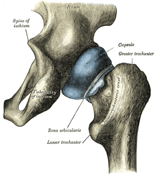
The capsule of hip joint, articular capsule, or capsular ligament is strong and dense attachment of the hip joint.

<span class="mw-page-title-main">Pelvis</span> Lower torso of the human body

The pelvis is the lower part of the trunk, between the abdomen and the thighs, together with its embedded skeleton.

References

PD-icon.svgThis article incorporates text in the public domain from page 245 of the 20th edition of Gray's Anatomy (1918)

  1. 1 2 3 4 5 Gray's anatomy : the anatomical basis of clinical practice. Susan Standring (Forty-second ed.). [New York]. 2021. p. 1362. ISBN   978-0-7020-7707-4. OCLC   1201341621.{{cite book}}: CS1 maint: location missing publisher (link) CS1 maint: others (link)
  2. Gray, Henry (1918). Gray's Anatomy (20th ed.). p. 245.
  3. Federle, Michael P.; Rosado-de-Christenson, Melissa L.; Raman, Siva P.; Carter, Brett W., eds. (2017-01-01), "Female Pelvic Floor", Imaging Anatomy: Chest, Abdomen, Pelvis (Second Edition), Elsevier, pp. 1050–1077, ISBN   978-0-323-47781-9 , retrieved 2021-01-22
  4. Khoury JG, Brandser EA, Found EM, Buckwalter JA (1998). "Non-traumatic lesser trochanter avulsion: a report of three cases". Iowa Orthop J. 18: 150–4. PMC   2378165 . PMID   9807723.
  5. Liu, J.; Olsen, P. (2010). "The Phylogenetic Relationships of Eucynodontia (Amniota: Synapsida)". Journal of Mammalian Evolution. 17 (3): 151. doi:10.1007/s10914-010-9136-8. S2CID   40871206.