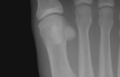
| First metatarsal bone | |
|---|---|
 The first metatarsal. (Left.) | |
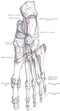
 Bones of the right foot. Dorsal surface. The first metatarsal bone is shown in yellow furthest to the left | |
| Details | |
| Articulations | Proximal phalange and medial cuneiform |
| Identifiers | |
| Latin | os metatarsale I |
| TA2 | 1500 |
| FMA | 24502 |
| Anatomical terms of bone | |
The first metatarsal bone is the bone in the foot just behind the big toe. The first metatarsal bone is the shortest of the metatarsal bones and by far the thickest and strongest of them. [1]
Contents
Like the four other metatarsals, it can be divided into three parts: base, body and head. The base is the part closest to the ankle and the head is closest to the big toe. The narrowed part in the middle is referred to as the body of the bone. The bone is somewhat flattened, giving it two sides: the plantar (towards the sole of the foot) and the dorsal side (the area facing upwards while standing). [1]
The base presents, as a rule, no articular facets (joint surfaces) on its sides, but occasionally on the lateral side there is an oval facet, by which it articulates with the second metatarsal. On the lateral part of the plantar surface there is a rough oval prominence, or tuberosity, for the insertion of the tendon of the fibularis longus.
The first metatarsal articulates (forms joints) with the medial cuneiform and to a small extent with the intermediate cuneiform bone. [2] Its proximal articular surface is large and kidney-shaped. Its circumference is grooved, for the tarsometatarsal ligaments, and medially gives insertion to part of the tendon of the tibialis anterior.
The body of the bone is strong, and of well-marked prismoid form.
The head is large; on its plantar surface are two grooved facets on which the sesamoid bones glide. The facets are separated by a smooth elevation.