
The foot is an anatomical structure found in many vertebrates. It is the terminal portion of a limb which bears weight and allows locomotion. In many animals with feet, the foot is a separate organ at the terminal part of the leg made up of one or more segments or bones, generally including claws and/or nails.

In the human body, the cuboid bone is one of the seven tarsal bones of the foot.

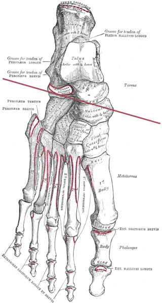
The metatarsal bones or metatarsus are a group of five long bones in the midfoot, located between the tarsal bones and the phalanges (toes). Lacking individual names, the metatarsal bones are numbered from the medial side : the first, second, third, fourth, and fifth metatarsal. The metatarsals are analogous to the metacarpal bones of the hand. The lengths of the metatarsal bones in humans are, in descending order, second, third, fourth, fifth, and first. A bovine hind leg has two metatarsals.

The ankle, the talocrural region or the jumping bone (informal) is the area where the foot and the leg meet. The ankle includes three joints: the ankle joint proper or talocrural joint, the subtalar joint, and the inferior tibiofibular joint. The movements produced at this joint are dorsiflexion and plantarflexion of the foot. In common usage, the term ankle refers exclusively to the ankle region. In medical terminology, "ankle" can refer broadly to the region or specifically to the talocrural joint.

In humans and many other primates, the calcaneus or heel bone is a bone of the tarsus of the foot which constitutes the heel. In some other animals, it is the point of the hock.

The navicular bone is a small bone found in the feet of most mammals.
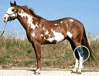
The hock, tarsus or uncommonly gambrel, is the region formed by the tarsal bones connecting the tibia and metatarsus of a digitigrade or unguligrade quadrupedal mammal, such as a horse, cat, or dog. This joint may include articulations between tarsal bones and the fibula in some species, while in others the fibula has been greatly reduced and is only found as a vestigial remnant fused to the distal portion of the tibia. It is the anatomical homologue of the ankle of the human foot. While homologous joints occur in other tetrapods, the term is generally restricted to mammals, particularly long-legged domesticated species.
The talus, talus bone, astragalus, or ankle bone is one of the group of foot bones known as the tarsus. The tarsus forms the lower part of the ankle joint. It transmits the entire weight of the body from the lower legs to the foot.
In human anatomy, the subtalar joint, also known as the talocalcaneal joint, is a joint of the foot. It occurs at the meeting point of the talus and the calcaneus.

The arches of the foot, formed by the tarsal and metatarsal bones, strengthened by ligaments and tendons, allow the foot to support the weight of the body in the erect posture with the least weight.

The talocalcaneonavicular joint is a ball and socket joint in the foot; the rounded head of the talus is received into the concavity formed by the posterior surface of the navicular, the anterior articular surface of the calcaneus, and the upper surface of the plantar calcaneonavicular ligament.

The bifurcated ligament is a strong band, attached behind to the deep hollow on the upper surface of the calcaneus and dividing in front in a Y-shaped manner into a calcaneocuboid and a calcaneonavicular part.
The transverse tarsal joint or midtarsal joint or Chopart's joint is formed by the articulation of the calcaneus with the cuboid, and the articulation of the talus with the navicular.

A calcaneal fracture is a break of the calcaneus. Symptoms may include pain, bruising, trouble walking, and deformity of the heel. It may be associated with breaks of the hip or back.
The Ponseti method is a manipulative technique that corrects congenital clubfoot without invasive surgery. It was developed by Ignacio V. Ponseti of the University of Iowa Hospitals and Clinics, US, in the 1950s, and was repopularized in 2000 by John Herzenberg in the US and Europe and in Africa by NHS surgeon Steve Mannion. It is a standard treatment for clubfoot.

Pigeon toe, also known as in-toeing, is a condition which causes the toes to point inward when walking. It is most common in infants and children under two years of age and, when not the result of simple muscle weakness, normally arises from underlying conditions, such as a twisted shin bone or an excessive anteversion resulting in the twisting of the thigh bone when the front part of a person's foot is turned in.
Chopart's fracture–dislocation is a dislocation of the mid-tarsal joints of the foot, often with associated fractures of the calcaneus, cuboid and navicular.

The calcaneal pitch is an angle used mainly in the diagnosis and severity grading of flat feet and pes cavus.

The sinus tarsi, also known as the talocalcaneal sulcus, is a cylindrical canal in the hindfoot. It has a complex anatomy, with five ligamentous structures and a pad of adipose tissue (fat). The tarsal canal opens up into the sinus tarsi, however, the tarsal canal is a distinct structure.
Subtalar arthroereisis is a common treatment for symptomatic pes planus, also known as flatfoot. There are two forms of pes planus: rigid flatfoot (RFF) and flexible flatfoot (FFF). The symptoms of the former typically necessitate surgical intervention. The latter may manifest fatigue or pain, but is typically asymptomatic.

 Foot bones - tarsus, metatarsus and phalanges.
 Bones of the right foot. Dorsal surface.
 Bones of the right foot. Plantar surface.
 CT 3D human Foot Skin and Bone
 Skeleton of foot. Medial aspect.
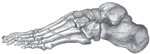 Skeleton of foot. Lateral aspect.
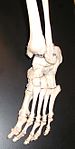 Bones of the feet from an actual skeleton.
 Skeleton of Manus and Pes of a Tailed Batrachian (from Professor Gegenbaur's "Tarsus and Carpus").
 Bones of foot
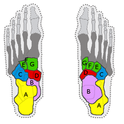


