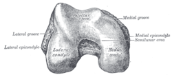
The humerus is a long bone in the arm that runs from the shoulder to the elbow. It connects the scapula and the two bones of the lower arm, the radius and ulna, and consists of three sections. The humeral upper extremity consists of a rounded head, a narrow neck, and two short processes. The body is cylindrical in its upper portion, and more prismatic below. The lower extremity consists of 2 epicondyles, 2 processes, and 3 fossae. As well as its true anatomical neck, the constriction below the greater and lesser tubercles of the humerus is referred to as its surgical neck due to its tendency to fracture, thus often becoming the focus of surgeons.

In anatomy, flexor carpi radialis is a muscle of the human forearm that acts to flex and (radially) abduct the hand. The Latin carpus means wrist; hence flexor carpi is a flexor of the wrist.

In human anatomy, the ulnar nerve is a nerve that runs near the ulna bone. The ulnar collateral ligament of elbow joint is in relation with the ulnar nerve. The nerve is the largest in the human body unprotected by muscle or bone, so injury is common. This nerve is directly connected to the little finger, and the adjacent half of the ring finger, innervating the palmar aspect of these fingers, including both front and back of the tips, perhaps as far back as the fingernail beds.

The ulnar collateral ligament (UCL) or internal lateral ligament is a thick triangular ligament at the medial aspect of the elbow uniting the distal aspect of the humerus to the proximal aspect of the ulna.

The cubital fossa, chelidon or inside of elbow is the area on the anterior side of the upper part between the arm and forearm of a human or other hormid animals. It lies anteriorly to the elbow when in standard anatomical position.

The lateral epicondyle of the humerus is a large, tuberculated eminence, curved a little forward, and giving attachment to the radial collateral ligament of the elbow joint, and to a tendon common to the origin of the supinator and some of the extensor muscles. Specifically, these extensor muscles include the anconeus muscle, the supinator, extensor carpi radialis brevis, extensor digitorum, extensor digiti minimi, and extensor carpi ulnaris. In birds, where the arm is somewhat rotated compared to other tetrapods, it is termed dorsal epicondyle of the humerus. In comparative anatomy, the term ectepicondyle is sometimes used.
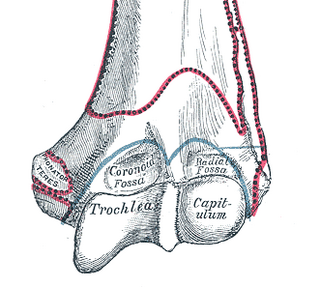
In the human arm, the humeral trochlea is the medial portion of the articular surface of the elbow joint which articulates with the trochlear notch on the ulna in the forearm.

The inferior ulnar collateral artery is an artery in the arm. It arises about 5 cm. above the elbow from the brachial artery.
The superior ulnar collateral artery, of small size, arises from the brachial artery a little below the middle of the arm; it frequently springs from the upper part of the a. profunda brachii.
The deep artery of arm is a large artery of the arm which arises from the brachial artery. It descends in the arm before ending by anastomosing with the radial recurrent artery.

The lower extremity of femur is the lower end of the femur in human and other animals, closer to the knee. It is larger than the upper extremity of femur, is somewhat cuboid in form, but its transverse diameter is greater than its antero-posterior; it consists of two oblong eminences known as the lateral condyle and medial condyle.

The medial epicondyle of the humerus is an epicondyle of the humerus bone of the upper arm in humans. It is larger and more prominent than the lateral epicondyle and is directed slightly more posteriorly in the anatomical position. In birds, where the arm is somewhat rotated compared to other tetrapods, it is called the ventral epicondyle of the humerus. In comparative anatomy, the more neutral term entepicondyle is used.

The medial epicondyle of the femur is an epicondyle, a bony protrusion, located on the medial side of the femur at its distal end.

The posterior ulnar recurrent artery is an artery in the forearm. It is one of two recurrent arteries that arises from the ulnar artery, the other being the anterior ulnar recurrent artery. The posterior ulnar recurrent artery being much larger than the anterior and also arises somewhat lower than it.
The brachial fascia is continuous with that covering the deltoideus and the pectoralis major muscle, by means of which it is attached, above, to the clavicle, acromion, and spine of the scapula; it forms a thin, loose, membranous sheath for the muscles of the arm, and sends septa between them; it is composed of fibers disposed in a circular or spiral direction, and connected together by vertical and oblique fibers.

The fascial compartments of arm refers to the specific anatomical term of the compartments within the upper segment of the upper limb of the body. The upper limb is divided into two segments, the arm and the forearm. Each of these segments is further divided into two compartments which are formed by deep fascia – tough connective tissue septa (walls). Each compartment encloses specific muscles and nerves.

One or two supratrochlear lymph nodes are placed above the medial epicondyle of the humerus, medial to the basilic vein.

The humeroulnar joint is part of the elbow-joint. It is composed of two bones, the humerus and ulna, and is the junction between the trochlear notch of ulna and the trochlea of humerus. It is classified as a simple hinge-joint, which allows for movements of flexion, extension and circumduction. Owing to the obliquity of the trochlea of the humerus, this movement does not take place in the antero-posterior plane of the body of the humerus.

The elbow is the region between the upper arm and the forearm that surrounds the elbow joint. The elbow includes prominent landmarks such as the olecranon, the cubital fossa, and the lateral and the medial epicondyles of the humerus. The elbow joint is a hinge joint between the arm and the forearm; more specifically between the humerus in the upper arm and the radius and ulna in the forearm which allows the forearm and hand to be moved towards and away from the body. The term elbow is specifically used for humans and other primates, and in other vertebrates it is not used. In those cases, forelimb plus joint is used.

Many anatomical terms descriptive of bone are defined in anatomical terminology, and are often derived from Greek and Latin. Bone in the human body is categorized into long bone, short bone, flat bone, irregular bone and sesamoid bone.