
In humans and other primates, the knee joins the thigh with the leg and consists of two joints: one between the femur and tibia, and one between the femur and patella. It is the largest joint in the human body. The knee is a modified hinge joint, which permits flexion and extension as well as slight internal and external rotation. The knee is vulnerable to injury and to the development of osteoarthritis.

The fibula or calf bone is a leg bone located on the lateral side of the tibia, with which it is connected above and below. It is the smaller of the two bones and in proportion to its length, the slenderest of all the long bones. Its upper extremity is small, placed toward the back of the head of the tibia, below the level of the knee joint, and excluded from the formation of this joint. Its lower extremity inclines a little forward, so as to be on a plane anterior to that of the upper end; it projects below the tibia, and forms the lateral part of the ankle-joint.

The inferior colliculus (IC) is the principal midbrain nucleus of the auditory pathway and receives input from several peripheral brainstem nuclei in the auditory pathway, as well as inputs from the auditory cortex. The inferior colliculus has three subdivisions: the central nucleus, a dorsal cortex by which it is surrounded, and an external cortex which is located laterally. Its bimodal neurons are implicated in auditory-somatosensory interaction, receiving projections from somatosensory nuclei. This multisensory integration may underlie a filtering of self-effected sounds from vocalization, chewing, or respiration activities.

The ulnar collateral ligament is a thick triangular band at the medial aspect of the elbow uniting the distal aspect of the humerus to the proximal aspect of the ulna.
The drawer test is used in the initial clinical assessment of suspected rupture of the cruciate ligaments in the knee. The patient should be supine with the hips flexed to 45 degrees, the knees flexed to 90 degrees and the feet flat on table. The examiner positions himself by sitting on the examination table in front of the involved knee and grasping the tibia just below the joint line of the knee. The thumbs are placed along the joint line on either side of the patellar tendon. The tibia is then drawn forward anteriorly. An increased amount of anterior tibial translation compared with the opposite limb or lack of a firm end-point may indicate either a sprain of the anteromedial bundle or complete tear of the ACL. If the tibia pulls forward or backward more than normal, the test is considered positive. Excessive displacement of the tibia anteriorly suggests that the ACL is injured, whereas excessive posterior displacement of the tibia may indicate injury of the posterior cruciate ligament.
The semimembranosus is the most medial of the three hamstring muscles. It is so named because it has a flat tendon of origin. It lies posteromedially in the thigh, deep to the semitendinosus.

An ankle fracture is a break of the ankle bones. It is typically diagnosed by X-ray. Treatment is with splinting, casting or surgery. In children ankle fractures occur in about 1 per 1000 per year.

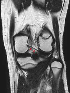
The medial meniscus is a fibrocartilage semicircular band that spans the knee joint medially, located between the medial condyle of the femur and the medial condyle of the tibia. It is also referred to as the internal semilunar fibrocartilage. The medial meniscus has more of a crescent shape while the lateral meniscus is more circular. The anterior aspects of both menisci are connected by the transverse ligament. It is a common site of injury, especially if the knee is twisted.
The lateral meniscus is a fibrocartilaginous band that spans the lateral side of the interior of the knee joint. It is one of two menisci of the knee, the other being the medial meniscus. It is nearly circular and covers a larger portion of the articular surface than the medial. It can occasionally be injured or torn by twisting the knee or applying direct force, as seen in contact sports.

The facial colliculus is an elevated area located on the pontine tegmentum in the floor of the fourth ventricle. It is formed by fibers from the facial motor nucleus of the facial nerve as they loop over the abducens nucleus. Thus a lesion to the facial colliculus would result in ipsilateral facial paralysis and ipsilateral unopposed eye medial deviation.

The lower extremity of the humerus is flattened from before backward, and curved slightly forward; it ends below in a broad, articular surface, which is divided into two parts by a slight ridge.

The patellar ligament is the distal portion of the common tendon of the quadriceps femoris, which is continued from the patella to the tibial tuberosity. It is also sometimes called the patellar tendon as it is a continuation of the quadriceps tendon.

A malleolus is the bony prominence on each side of the human ankle.

The coronary ligaments of the knee are portions of the joint capsule which connect the inferior edges of the fibrocartilaginous menisci to the periphery of the tibial plateaus.
The Posterior meniscofemoral ligament is a small fibrous band of the knee joint. It attaches to the posterior area of the lateral meniscus and crosses superiorly and medially behind the posterior cruciate ligament to attach to the medial condyle of the femur.

The intercondylar area is the separation between the medial and lateral condyle on the upper extremity of the tibia. The anterior and posterior cruciate ligaments and the menisci attach to the intercondylar area.
The anterior colliculus is the anterior portion of the medial malleolus of the distal tibia, forming part of the ankle mortise. It has an attachment of the anterior tibiotalar ligament.

