The leg is the entire lower limb of the human body, including the foot, thigh or sometimes even the hip or buttock region. The major bones of the leg are the femur, tibia, and adjacent fibula. The thigh is between the hip and knee, while the calf (rear) and shin (front) are between the knee and foot.

In humans and other primates, the knee joins the thigh with the leg and consists of two joints: one between the femur and tibia, and one between the femur and patella. It is the largest joint in the human body. The knee is a modified hinge joint, which permits flexion and extension as well as slight internal and external rotation. The knee is vulnerable to injury and to the development of osteoarthritis.

In human anatomy, a hamstring is any one of the three posterior thigh muscles between the hip and the knee.

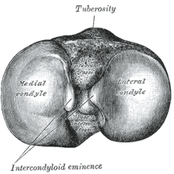
The tibia, also known as the shinbone or shankbone, is the larger, stronger, and anterior (frontal) of the two bones in the leg below the knee in vertebrates ; it connects the knee with the ankle. The tibia is found on the medial side of the leg next to the fibula and closer to the median plane. The tibia is connected to the fibula by the interosseous membrane of leg, forming a type of fibrous joint called a syndesmosis with very little movement. The tibia is named for the flute tibia. It is the second largest bone in the human body, after the femur. The leg bones are the strongest long bones as they support the rest of the body.

The patella, also known as the kneecap, is a flat, rounded triangular bone which articulates with the femur and covers and protects the anterior articular surface of the knee joint. The patella is found in many tetrapods, such as mice, cats, birds and dogs, but not in whales, or most reptiles.

The quadriceps femoris muscle is a large muscle group that includes the four prevailing muscles on the front of the thigh. It is the sole extensor muscle of the knee, forming a large fleshy mass which covers the front and sides of the femur. The name derives from Latin four-headed muscle of the femur.
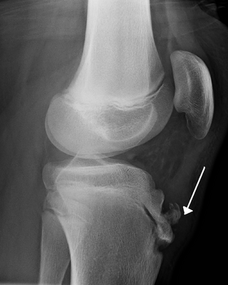
Osgood–Schlatter disease (OSD) is inflammation of the patellar ligament at the tibial tuberosity (apophysitis). It is characterized by a painful bump just below the knee that is worse with activity and better with rest. Episodes of pain typically last a few weeks to months. One or both knees may be affected and flares may recur.

The vastus medialis is an extensor muscle located medially in the thigh that extends the knee. The vastus medialis is part of the quadriceps muscle group.
The vastus lateralis, also called the vastus externus, is the largest and most powerful part of the quadriceps femoris, a muscle in the thigh. Together with other muscles of the quadriceps group, it serves to extend the knee joint, moving the lower leg forward. It arises from a series of flat, broad tendons attached to the femur, and attaches to the outer border of the patella. It ultimately joins with the other muscles that make up the quadriceps in the quadriceps tendon, which travels over the knee to connect to the tibia. The vastus lateralis is the recommended site for intramuscular injection in infants less than 7 months old and those unable to walk, with loss of muscular tone.
The semimembranosus muscle is the most medial of the three hamstring muscles in the thigh. It is so named because it has a flat tendon of origin. It lies posteromedially in the thigh, deep to the semitendinosus muscle. It extends the hip joint and flexes the knee joint.

The semitendinosus is a long superficial muscle in the back of the thigh. It is so named because it has a very long tendon of insertion. It lies posteromedially in the thigh, superficial to the semimembranosus.
The rectus femoris muscle is one of the four quadriceps muscles of the human body. The others are the vastus medialis, the vastus intermedius, and the vastus lateralis. All four parts of the quadriceps muscle attach to the patella by the quadriceps tendon.
The knee examination, in medicine and physiotherapy, is performed as part of a physical examination, or when a patient presents with knee pain or a history that suggests a pathology of the knee joint.

The patellar tendon is the distal portion of the common tendon of the quadriceps femoris, which is continued from the patella to the tibial tuberosity. It is also sometimes called the patellar ligament as it forms a bone to bone connection when the patella is fully ossified.

The articular capsule of the knee joint is the wide and lax joint capsule of the knee. It is thin in front and at the side, and contains the patella, ligaments, menisci, and bursae of the knee. The capsule consists of an inner synovial membrane, and an outer fibrous membrane separated by fatty deposits anteriorly and posteriorly.
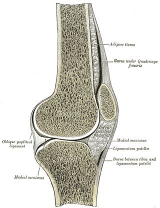
The knee bursae are the fluid-filled sacs and synovial pockets that surround and sometimes communicate with the knee joint cavity. The bursae are thin-walled, and filled with synovial fluid. They represent the weak point of the joint, but also provide enlargements to the joint space. They can be grouped into either communicating and non-communicating bursae or, after their location – frontal, lateral, or medial.

A patellar dislocation is a knee injury in which the patella (kneecap) slips out of its normal position. Often the knee is partly bent, painful and swollen. The patella is also often felt and seen out of place. Complications may include a patella fracture or arthritis.
The vastus muscles are three of the four muscles that make up the quadriceps femoris muscle of the thigh. The three muscles are the vastus intermedius, the vastus lateralis, and the vastus medialis located in the middle, on the outside, and inside of the thigh, respectively. The fourth muscle is the rectus femoris muscle a large fleshy muscle which covers the front and sides of the femur.
Medial knee injuries are the most common type of knee injury. The medial ligament complex of the knee consists of:
In the skeleton of humans and other animals, a tubercle, tuberosity or apophysis is a protrusion or eminence that serves as an attachment for skeletal muscles. The muscles attach by tendons, where the enthesis is the connective tissue between the tendon and bone. A tuberosity is generally a larger tubercle.