In vertebrate anatomy, the pituitary gland, or hypophysis, is an endocrine gland about the size of a pea and weighing 0.5 grams (0.018 oz) in humans. It is a protrusion off the bottom of the hypothalamus at the base of the brain. The hypophysis rests upon the hypophysial fossa of the sphenoid bone in the center of the middle cranial fossa and is surrounded by a small bony cavity covered by a dural fold. The anterior pituitary is a lobe of the gland that regulates several physiological processes. The intermediate lobe synthesizes and secretes melanocyte-stimulating hormone. The posterior pituitary is a lobe of the gland that is functionally connected to the hypothalamus by the median eminence via a small tube called the pituitary stalk.

Standard anatomical terms of location deal unambiguously with the anatomy of animals, including humans.
The forearm is the region of the upper limb between the elbow and the wrist. The term forearm is used in anatomy to distinguish it from the arm, a word which is most often used to describe the entire appendage of the upper limb, but which in anatomy, technically, means only the region of the upper arm, whereas the lower "arm" is called the forearm. It is homologous to the region of the leg that lies between the knee and the ankle joints, the crus.

The circle of Willis is a circulatory anastomosis that supplies blood to the brain and surrounding structures. It is named after Thomas Willis (1621–1675), an English physician.
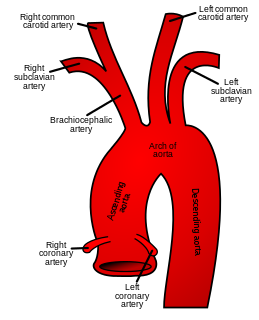
In human anatomy, the subclavian arteries are paired major arteries of the upper thorax, below the clavicle. They receive blood from the aortic arch. The left subclavian artery supplies blood to the left arm and the right subclavian artery supplies blood to the right arm, with some branches supplying the head and thorax. On the left side of the body, the subclavian comes directly off the aortic arch, while on the right side it arises from the relatively short brachiocephalic artery when it bifurcates into the subclavian and the right common carotid artery.

The mandibular nerve (V3) is the largest of the three divisions of the trigeminal nerve, the fifth cranial nerve (CN V).
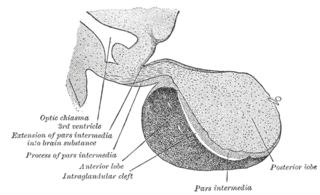
A major organ of the endocrine system, the anterior pituitary, is the glandular, anterior lobe that together with the posterior lobe makes up the pituitary gland (hypophysis). The anterior pituitary regulates several physiological processes including stress, growth, reproduction and lactation. Proper functioning of the anterior pituitary and of the organs it regulates can often be ascertained via blood tests that measure hormone levels.

The cingulate cortex is a part of the brain situated in the medial aspect of the cerebral cortex. The cingulate cortex includes the entire cingulate gyrus, which lies immediately above the corpus callosum, and the continuation of this in the cingulate sulcus. The cingulate cortex is usually considered part of the limbic lobe.

The deltoid muscle is the muscle forming the rounded contour of the human shoulder. It is also known as the 'common shoulder muscle', particularly in other animals such as the domestic cat. Anatomically, it appears to be made up of three distinct sets of fibers though electromyography suggests that it consists of at least seven groups that can be independently coordinated by the nervous system.

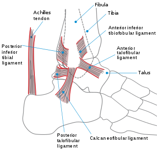
The anterior cruciate ligament (ACL) is one of a pair of cruciate ligaments in the human knee. The two ligaments are also called cruciform ligaments, as they are arranged in a crossed formation. In the quadruped stifle joint, based on its anatomical position, it is also referred to as the cranial cruciate ligament. The term cruciate translates to cross. This name is fitting because the ACL crosses the posterior cruciate ligament to form an “X”. It is composed of strong fibrous material and assists in controlling excessive motion. This is done by limiting mobility of the joint. The anterior cruciate ligament is one of the four main ligaments of the knee, providing 85% of the restraining force to anterior tibial displacement at 30 degrees and 90 degrees of knee flexion. The ACL is the most injured ligament of the four located in the knee.

The tibialis anterior is a muscle in humans that originates in the upper two-thirds of the lateral (outside) surface of the tibia and inserts into the medial cuneiform and first metatarsal bones of the foot. It acts to dorsiflex and invert the foot. This muscle is mostly located near the shin.
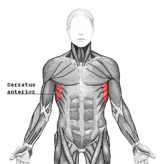
The serratus anterior is a muscle that originates on the surface of the 1st to 8th ribs at the side of the chest and inserts along the entire anterior length of the medial border of the scapula. The serratus anterior acts to pull the scapula forward around the thorax. The muscle is named from Latin: serrare = to saw, referring to the shape, anterior = on the front side of the body.

The scalene muscles are a group of three pairs of muscles in the lateral neck, namely the anterior scalene, middle scalene, and posterior scalene. They are innervated by the fourth, fifth, and sixth cervical spinal nerves (C4-C6).

The external oblique muscle is the largest and the outermost of the three flat muscles of the lateral anterior abdomen.

The anterior chamber (AC) is the aqueous humor-filled space inside the eye between the iris and the cornea's innermost surface, the endothelium. Hyphema, anterior uveitis and glaucoma are three main pathologies in this area. In hyphema, blood fills the anterior chamber as a result of a hemorrhage, most commonly after a blunt eye injury. Anterior uveitis is an inflammatory process affecting the iris and ciliary body, with resulting inflammatory signs in the anterior chamber. In glaucoma, blockage of the trabecular meshwork prevents the normal outflow of aqueous humour, resulting in increased intraocular pressure, progressive damage to the optic nerve head, and eventually blindness.

The anterior grey column is the front column of grey matter in the spinal cord. It is one of the three grey columns. The anterior grey column contains motor neurons that affect the skeletal muscles while the posterior grey column receives information regarding touch and sensation. The anterior grey column is the column where the cell bodies of alpha motor neurons are located.
Anterior cruciate ligament injury is when the anterior cruciate ligament (ACL) is either stretched, partially torn, or completely torn. The most common injury is a complete tear. Symptoms include pain, a popping sound during injury, instability of the knee, and joint swelling. Swelling generally appears within a couple of hours. In approximately 50% of cases, other structures of the knee such as surrounding ligaments, cartilage, or meniscus are damaged.

The spinal cord is a long, thin, tubular structure made up of nervous tissue, that extends from the medulla oblongata in the brainstem to the lumbar region of the vertebral column. It encloses the central canal of the spinal cord that contains cerebrospinal fluid. The brain and spinal cord together make up the central nervous system (CNS). In humans, the spinal cord begins at the occipital bone where it passes through the foramen magnum, and meets and enters the spinal canal at the beginning of the cervical vertebrae. The spinal cord extends down to between the first and second lumbar vertebrae where it ends. The enclosing bony vertebral column protects the relatively shorter spinal cord. It is around 45 cm (18 in) in men and around 43 cm (17 in) long in women. Also, the spinal cord has a varying width, ranging from 13 mm thick in the cervical and lumbar regions to 6.4 mm thick in the thoracic area.

The pelvis is either the lower part of the trunk of the human body between the abdomen and the thighs or the skeleton embedded in it.