Metatarsal bones

Last updated
Metatarsal bones
Metatarsal bones01 - superior view.png
Skeleton of foot. Superior view. Metatarsals shown in green
Gray291 - Metatarsus.png
Skeleton of left foot. Lateral aspect. Metatarsals shown in purple
Details
Identifiers
Latin metatarsus
pl. ossa metatarsi (also: ossa metatarsalia)
MeSH D008682
TA98 A02.5.17.001
TA2 1495
FMA 71340
Anatomical terms of bone

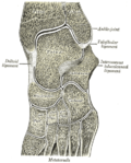
The metatarsal bones or metatarsus (pl.: metatarsi) are a group of five long bones in the midfoot, located between the tarsal bones (which forms the heel and the ankle) and the phalanges (toes). Lacking individual names, the metatarsal bones are numbered from the medial side (the side of the great toe): the first, second, third, fourth, and fifth metatarsal (often depicted with Roman numerals). The metatarsals are analogous to the metacarpal bones of the hand. The lengths of the metatarsal bones in humans are, in descending order, second, third, fourth, fifth, and first. [1] A bovine hind leg has two metatarsals. [2]

Contents

Structure

The five metatarsals are dorsal convex long bones consisting of a shaft or body, a base (proximally), and a head (distally). [3] The body is prismoid in form, tapers gradually from the tarsal to the phalangeal extremity, and is curved longitudinally, so as to be concave below, slightly convex above. The base or posterior extremity is wedge-shaped, articulating proximally with the tarsal bones, and by its sides with the contiguous metatarsal bones: its dorsal and plantar surfaces are rough for the attachment of ligaments. The head or distal extremity presents a convex articular surface, oblong from above downward, and extending farther backward below than above. Its sides are flattened, and on each is a depression, surmounted by a tubercle, for ligamentous attachment. Its plantar surface is grooved antero-posteriorly for the passage of the flexor tendons, and marked on either side by an articular eminence continuous with the terminal articular surface. [4]

During growth, the growth plates are located distally on the metatarsals, except on the first metatarsal where it is located proximally. Yet it is quite common to have an accessory growth plate on the distal first metatarsal. [5]

Articulations

Bones of the right foot. Dorsal surface. Metatarsus shown in yellow. Ospied-en.svg
Bones of the right foot. Dorsal surface. Metatarsus shown in yellow.

The base of each metatarsal bone articulates with one or more of the tarsal bones at the tarsometatarsal joints, and the head with one of the first row of phalanges at the metatarsophalangeal joints. Their bases also articulate with each other at the intermetatarsal joints

Muscle attachments

Muscle attachments (seen from above) Gray268 - Mratatarsus.png
Muscle attachments (seen from above)
Muscle attachments (seen from below) Gray269 - Mratatarsus.png
Muscle attachments (seen from below)
MuscleDirectionAttachment [7]
Tibialis anterior InsertionBasis of first metatarsal
Peroneous tertius InsertionDorsal side basis of fifth metatarsal
Peroneous longus InsertionTuberosity of first metatarsal
Peroneous brevis InsertionTuberosity of fifth metatarsal
Horizontal head of adductor hallucis OriginDeep transverse metatarsal ligament
Flexor digiti minimi brevis OriginBasis of fifth metatarsal
Plantar interossei OriginMedial side of third, fourth and fifth metatarsal
Dorsal interossei Origin First to fifth metatarsal

Clinical significance

Injuries

Fracture of the base of the 5th metatarsal FractureBaseof5th.png
Fracture of the base of the 5th metatarsal

The metatarsal bones are often broken by association football (soccer) players. These and other recent cases have been attributed to the lightweight design of modern football boots, which provide less protection to the foot. In 2010 some football players began testing a new sock that incorporated a rubber silicone pad over the foot to provide protection to the top of the foot. [8] Stress fractures are thought to account for 16% of injuries related to sports participation, and the metatarsals are the bones most often involved. These fractures are sometimes called march fractures, based on their traditional association with military recruits after long marches. The second and third metatarsals are fixed while walking, thus these metatarsals are common sites of injury. The fifth metatarsal may be fractured if the foot is oversupinated during locomotion. [9]

Protection from injuries can be given by the use of safety footwear which can use built-in or removable metatarsal guards.

Additional images

See also

Notes

  1. Bojsen-Møller, Finn; Simonsen, Erik B.; Tranum-Jensen, Jørgen (2001). Bevægeapparatets anatomi[Anatomy of the Locomotive Apparatus] (in Danish) (12th ed.). p. 246. ISBN   978-87-628-0307-7.
  2. "Identification – cattle hock bone |".
  3. Platzer 2004, p. 220
  4. Gray's 1918, 6d. 2. The Metatarsus
  5. Mathis, SK; Frame, BA; Smith, CE (1989). "Distal first metatarsal epiphysis. A common pediatric variant". Journal of the American Podiatric Medical Association. 79 (8): 375–79. doi:10.7547/87507315-79-8-375. ISSN   8750-7315. PMID   2681682.
  6. 1 2 3 4 5 Platzer 2004, p. 218
  7. Bojsen-Møller, Finn; Simonsen, Erik B.; Tranum-Jensen, Jørgen (2001). Bevægeapparatets anatomi[Anatomy of the Locomotive Apparatus] (in Danish) (12th ed.). pp. 364–67. ISBN   978-87-628-0307-7.
  8. Bill, Mills (11 December 2010). "Sock boffs may have cured metatarsal woes for Rooney and Co". www.mirrorfootball.co.uk. Retrieved 12 December 2010.
  9. Perron, Andrew D. (2005-11-23). "Metatarsal Stress Fracture" . Retrieved 2007-09-13.

Related Research Articles

<span class="mw-page-title-main">Foot</span> Anatomical structure found in vertebrates

The foot is an anatomical structure found in many vertebrates. It is the terminal portion of a limb which bears weight and allows locomotion. In many animals with feet, the foot is a separate organ at the terminal part of the leg made up of one or more segments or bones, generally including claws and/or nails.

<span class="mw-page-title-main">Human leg</span> Lower extremity or limb of the human body (foot, lower leg, thigh and hip)

The leg is the entire lower limb of the human body, including the foot, thigh or sometimes even the hip or buttock region. The major bones of the leg are the femur, tibia, and adjacent fibula. The thigh is between the hip and knee, while the calf (rear) and shin (front) are between the knee and foot.

<span class="mw-page-title-main">Femur</span> Thigh bone

The femur, or thigh bone is the only bone in the thigh. The thigh is the region of the lower limb between the hip and the knee. In many four-legged animals the femur is the upper bone of the hindleg.

<span class="mw-page-title-main">Cuboid bone</span> Bone of the ankle

In the human body, the cuboid bone is one of the seven tarsal bones of the foot.

<span class="mw-page-title-main">Tibia</span> Leg bone in vertebrates

The tibia, also known as the shinbone or shankbone, is the larger, stronger, and anterior (frontal) of the two bones in the leg below the knee in vertebrates ; it connects the knee with the ankle. The tibia is found on the medial side of the leg next to the fibula and closer to the median plane. The tibia is connected to the fibula by the interosseous membrane of leg, forming a type of fibrous joint called a syndesmosis with very little movement. The tibia is named for the flute tibia. It is the second largest bone in the human body, after the femur. The leg bones are the strongest long bones as they support the rest of the body.

<span class="mw-page-title-main">Fibula</span> Leg bone in vertebrates

The fibula or calf bone is a leg bone on the lateral side of the tibia, to which it is connected above and below. It is the smaller of the two bones and, in proportion to its length, the most slender of all the long bones. Its upper extremity is small, placed toward the back of the head of the tibia, below the knee joint and excluded from the formation of this joint. Its lower extremity inclines a little forward, so as to be on a plane anterior to that of the upper end; it projects below the tibia and forms the lateral part of the ankle joint.

<span class="mw-page-title-main">Metacarpal bones</span> Bones of hand

In human anatomy, the metacarpal bones or metacarpus, also known as the "palm bones", are the appendicular bones that form the intermediate part of the hand between the phalanges (fingers) and the carpal bones, which articulate with the forearm. The metacarpal bones are homologous to the metatarsal bones in the foot.

<span class="mw-page-title-main">Cuneiform bones</span> Three bones in the human foot

There are three cuneiform ("wedge-shaped") bones in the human foot:

<span class="mw-page-title-main">Calcaneus</span> Bone of the tarsus of the foot

In humans and many other primates, the calcaneus or heel bone is a bone of the tarsus of the foot which constitutes the heel. In some other animals, it is the point of the hock.

<span class="mw-page-title-main">Navicular bone</span> Small bone found in the feet of most mammals

The navicular bone is a small bone found in the feet of most mammals.

<span class="mw-page-title-main">Phalanx bone</span> Digital bone in the hands and feet of most vertebrates

The phalanges are digital bones in the hands and feet of most vertebrates. In primates, the thumbs and big toes have two phalanges while the other digits have three phalanges. The phalanges are classed as long bones.

<span class="mw-page-title-main">Tarsus (skeleton)</span> Bones of the foot

In the human body, the tarsus is a cluster of seven articulating bones in each foot situated between the lower end of the tibia and the fibula of the lower leg and the metatarsus. It is made up of the midfoot and hindfoot.

<span class="mw-page-title-main">Talus bone</span> One of the foot bones that forms the tarsus

The talus, talus bone, astragalus, or ankle bone is one of the group of foot bones known as the tarsus. The tarsus forms the lower part of the ankle joint. It transmits the entire weight of the body from the lower legs to the foot.

<span class="mw-page-title-main">Tarsometatarsal joints</span> Part of the human foot

The tarsometatarsal joints are arthrodial joints in the foot. The tarsometatarsal joints involve the first, second and third cuneiform bones, the cuboid bone and the metatarsal bones. The eponym of Lisfranc joint is 18th–19th-century surgeon and gynecologist Jacques Lisfranc de St. Martin.

<span class="mw-page-title-main">Interphalangeal joints of the foot</span>

The interphalangeal joints of the foot are between the phalanx bones of the toes in the feet.

<span class="mw-page-title-main">Fifth metatarsal bone</span>

The fifth metatarsal bone is a long bone in the foot, and is palpable along the distal outer edges of the feet. It is the second smallest of the five metatarsal bones. The fifth metatarsal is analogous to the fifth metacarpal bone in the hand.

<span class="mw-page-title-main">Fourth metatarsal bone</span>

The fourth metatarsal bone is a long bone in the foot. It is smaller in size than the third metatarsal bone and is the third longest of the five metatarsal bones. The fourth metatarsal is analogous to the fourth metacarpal bone in the hand

<span class="mw-page-title-main">Third metatarsal bone</span>

The third metatarsal bone is a long bone in the foot. It is the second longest metatarsal. The longest being the second metatarsal. The third metatarsal is analogous to the third metacarpal bone in the hand

<span class="mw-page-title-main">Second metatarsal bone</span>

The second metatarsal bone is a long bone in the foot. It is the longest of the metatarsal bones, being prolonged backward and held firmly into the recess formed by the three cuneiform bones. The second metatarsal forms joints with the second proximal phalanx through the metatarsophalangeal joint, the cuneiform bones, third metatarsal and occasionally the first metatarsal bone.

<span class="mw-page-title-main">First metatarsal bone</span>

The first metatarsal bone is the bone in the foot just behind the big toe. The first metatarsal bone is the shortest of the metatarsal bones and by far the thickest and strongest of them.

References