Dorsal interossei of the foot

Last updated
Dorsal interossei muscles
Gray446.png
The Interossei dorsales. Left foot.
Details
Origin metatarsals
Insertion proximal phalanges
Nerve lateral plantar nerve
Actions abduct toes
Antagonist Plantar interossei muscles
Identifiers
Latin musculi interossei dorsales pedis
TA98 A04.7.02.070
TA2 2686
FMA 37457
Anatomical terms of muscle

In human anatomy, the dorsal interossei of the foot are four muscles situated between the metatarsal bones.

Contents

Origin

The four interossei muscles are bipenniform muscles each originating by two heads from the proximal half of the sides of adjacent metatarsal bones. [1]

Insertion

The two heads of each muscle form a central tendon which passes forwards deep to the deep transverse metatarsal ligament. [1] The tendons are inserted on the bases of the second, third, and fourth proximal phalanges [2] and into the aponeurosis of the tendons of the extensor digitorum longus [3] without attaching to the extensor hoods of the toes. [1]

Thus, the first is inserted into the medial side of the second toe; the other three are inserted into the lateral sides of the second, third, and fourth toes. [3]

Action

The dorsal interossei abduct at the metatarsophalangeal joints of the third and fourth toes. Because there is a pair of dorsal interossei muscles attached on both sides of the second toe, simultaneous contraction of these muscles results in no movement. This arrangement of dorsal interossei makes the second toe the midline of the foot, whereas the midline of the hand (marked by dorsal interossei of hand) is in the third finger. [2]

Abduction is of little importance in the foot, but, together with the plantar interossei, the dorsal interossei also produce flexion at the metatarsophalangeal joints. Although small, the dorsal interossei are powerful muscles that, together with their plantar counterparts, control the direction of the toes during violent activity, thus allowing the long and short flexors to perform their actions. [1]

Because of the relationship to the metatarsophalangeal joints, the interossei muscles also contribute to maintaining the anterior metatarsal arch of the foot and also, to a limited extent, the medial and lateral longitudinal arches of the foot. [1]

Innervation

All dorsal interossei are innervated by the lateral plantar nerve (S2–3). Those in the fourth interosseous space are innervated by the superficial branch and the other by the deep branch. [1] The first and second dorsal interossei muscles additionally receive innervation from the lateral branch of the deep fibular nerve. [4]

Relations

In the angular interval left between the heads of each of the three lateral muscles, one of the perforating arteries passes to the dorsum of the foot; through the space between the heads of the first muscle the deep plantar branch of the dorsalis pedis artery enters the sole of the foot. [3]

See also

Additional images

Notes

PD-icon.svgThis article incorporates text in the public domain from page 495 of the 20th edition of Gray's Anatomy (1918)

  1. 1 2 3 4 5 6 Palastanga, Field & Soames 2006 , p. 311
  2. 1 2 Behnke 2006 , p. 227
  3. 1 2 3 Gray's Anatomy, 1918 (see infobox)
  4. Drake, Richard (2015). Gray's Anatomy for Students. Elsevier. ISBN   978-0702051333.

Related Research Articles

<span class="mw-page-title-main">Foot</span> Anatomical structure found in vertebrates

The foot is an anatomical structure found in many vertebrates. It is the terminal portion of a limb which bears weight and allows locomotion. In many animals with feet, the foot is a separate organ at the terminal part of the leg made up of one or more segments or bones, generally including claws and/or nails.

<span class="mw-page-title-main">Human leg</span> Lower extremity or limb of the human body (foot, lower leg, thigh and hip)

The leg is the entire lower limb of the human body, including the foot, thigh or sometimes even the hip or buttock region. The major bones of the leg are the femur, tibia, and adjacent fibula. The thigh is between the hip and knee, while the calf (rear) and shin (front) are between the knee and foot.

<span class="mw-page-title-main">Extensor hallucis longus muscle</span> Thin muscle, situated between the tibialis anterior and the extensor digitorum longus

The extensor hallucis longus muscle is a thin skeletal muscle, situated between the tibialis anterior and the extensor digitorum longus. It extends the big toe and dorsiflects the foot. It also assists with foot eversion and inversion.

<span class="mw-page-title-main">Tibialis anterior muscle</span> Flexor muscle in humans that dorsiflexes the foot

The tibialis anterior muscle is a muscle of the anterior compartment of the lower leg. It originates from the upper portion of the tibia; it inserts into the medial cuneiform and first metatarsal bones of the foot. It acts to dorsiflex and invert the foot. This muscle is mostly located near the shin.

<span class="mw-page-title-main">Extensor digitorum muscle</span> Muscle of the posterior forearm

The extensor digitorum muscle is a muscle of the posterior forearm present in humans and other animals. It extends the medial four digits of the hand. Extensor digitorum is innervated by the posterior interosseous nerve, which is a branch of the radial nerve.

<span class="mw-page-title-main">Palmar interossei muscles</span> Muscles between the metacarpals

In human anatomy, the palmar or volar interossei are four muscles, one on the thumb that is occasionally missing, and three small, unipennate, central muscles in the hand that lie between the metacarpal bones and are attached to the index, ring, and little fingers. They are smaller than the dorsal interossei of the hand.

<span class="mw-page-title-main">Extensor digitorum longus muscle</span> Pennate muscle, situated at the lateral part of the front of the leg

The extensor digitorum longus is a pennate muscle, situated at the lateral part of the front of the leg.

<span class="mw-page-title-main">Extensor digitorum brevis muscle</span> Muscle on the upper surface of the foot that helps extend digits 2 through 4

The extensor digitorum brevis muscle is a muscle on the upper surface of the foot that helps extend digits 2 through 4.
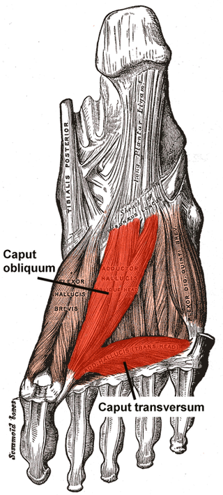
<span class="mw-page-title-main">Flexor hallucis brevis muscle</span> Muscle in sole of the foot that leads to the big toe

Flexor hallucis brevis muscle is a muscle of the foot that flexes the big toe.

<span class="mw-page-title-main">Plantar interossei muscles</span> Three muscles located between the metatarsal bones in the foot

In human anatomy, plantar interossei muscles are three muscles located between the metatarsal bones in the foot.

<span class="mw-page-title-main">Lumbricals of the foot</span> Four small skeletal muscles

The lumbricals are four small skeletal muscles, accessory to the tendons of the flexor digitorum longus muscle. They are numbered from the medial side of the foot.

<span class="mw-page-title-main">Deep fibular nerve</span> Type of nerve

The deep fibular nerve begins at the bifurcation of the common fibular nerve between the fibula and upper part of the fibularis longus, passes infero-medially, deep to the extensor digitorum longus, to the anterior surface of the interosseous membrane, and comes into relation with the anterior tibial artery above the middle of the leg; it then descends with the artery to the front of the ankle-joint, where it divides into a lateral and a medial terminal branch.

<span class="mw-page-title-main">Abductor digiti minimi muscle of foot</span> Muscle which lies along the lateral (outer) border of the foot

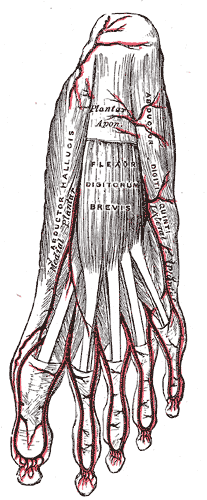
The abductor digiti minimi is a muscle which lies along the lateral (outer) border of the foot, and is in relation by its medial margin with the lateral plantar artery, vein and nerves.

<span class="mw-page-title-main">Dorsal interossei of the hand</span> Muscles between the metacarpals

In human anatomy, the dorsal interossei (DI) are four muscles in the back of the hand that act to abduct (spread) the index, middle, and ring fingers away from hand's midline and assist in flexion at the metacarpophalangeal joints and extension at the interphalangeal joints of the index, middle and ring fingers.

<span class="mw-page-title-main">Sole (foot)</span> Bottom part of foot

The sole is the bottom of the foot.

<span class="mw-page-title-main">Interphalangeal joints of the foot</span>

The interphalangeal joints of the foot are between the phalanx bones of the toes in the feet.

<span class="mw-page-title-main">Fifth metatarsal bone</span>

The fifth metatarsal bone is a long bone in the foot, and is palpable along the distal outer edges of the feet. It is the second smallest of the five metatarsal bones. The fifth metatarsal is analogous to the fifth metacarpal bone in the hand.

<span class="mw-page-title-main">Fourth metatarsal bone</span>

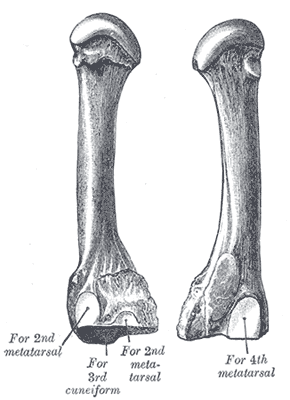
The fourth metatarsal bone is a long bone in the foot. It is smaller in size than the third metatarsal bone and is the third longest of the five metatarsal bones. The fourth metatarsal is analogous to the fourth metacarpal bone in the hand

<span class="mw-page-title-main">Third metatarsal bone</span>

The third metatarsal bone is a long bone in the foot. It is the second longest metatarsal. The longest being the second metatarsal. The third metatarsal is analogous to the third metacarpal bone in the hand

In the human foot, the plantar or volar plates are fibrocartilaginous structures found in the metatarsophalangeal (MTP) and interphalangeal (IP) joints. The anatomy and composition of the plantar plates are similar to the palmar plates in the metacarpophalangeal (MCP) and interphalangeal joints in the hand; the proximal origin is thin but the distal insertion is stout. Due to the weight-bearing nature of the human foot, the plantar plates are exposed to extension forces not present in the human hand.

References