
The foot is an anatomical structure found in many vertebrates. It is the terminal portion of a limb which bears weight and allows locomotion. In many animals with feet, the foot is a separate organ at the terminal part of the leg made up of one or more segments or bones, generally including claws and/or nails.

The leg is the entire lower limb of the human body, including the foot, thigh or sometimes even the hip or buttock region. The major bones of the leg are the femur, tibia, and adjacent fibula.
The flexor digitorum profundus or flexor digitorum communis profundus is a muscle in the forearm of humans that flexes the fingers. It is considered an extrinsic hand muscle because it acts on the hand while its muscle belly is located in the forearm.

The upper limbs or upper extremities are the forelimbs of an upright-postured tetrapod vertebrate, extending from the scapulae and clavicles down to and including the digits, including all the musculatures and ligaments involved with the shoulder, elbow, wrist and knuckle joints. In humans, each upper limb is divided into the shoulder, arm, elbow, forearm, wrist and hand, and is primarily used for climbing, lifting and manipulating objects. In anatomy, just as arm refers to the upper arm, leg refers to the lower leg.

The tibial nerve is a branch of the sciatic nerve. The tibial nerve passes through the popliteal fossa to pass below the arch of soleus.

The extensor digitorum muscle is a muscle of the posterior forearm present in humans and other animals. It extends the medial four digits of the hand. Extensor digitorum is innervated by the posterior interosseous nerve, which is a branch of the radial nerve.

In human anatomy, the dorsal interossei of the foot are four muscles situated between the metatarsal bones.

The flexor hallucis longus muscle (FHL) attaches to the plantar surface of phalanx of the great toe and is responsible for flexing that toe. The FHL is one of the three deep muscles of the posterior compartment of the leg, the others being the flexor digitorum longus and the tibialis posterior. The tibialis posterior is the most powerful of these deep muscles. All three muscles are innervated by the tibial nerve which comprises half of the sciatic nerve.

The flexor digitorum longus muscle or flexor digitorum communis longus is situated on the tibial side of the leg. At its origin it is thin and pointed, but it gradually increases in size as it descends. It serves to flex the second, third, fourth, and fifth toes.

The extensor digitorum longus is a pennate muscle, situated at the lateral part of the front of the leg.
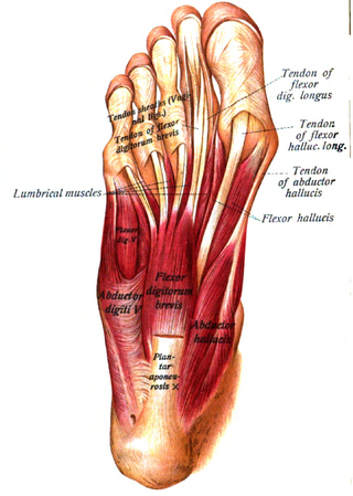
The abductor hallucis muscle is an intrinsic muscle of the foot. It participates in the abduction and flexion of the great toe.
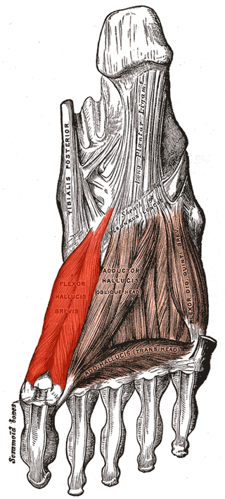
Flexor hallucis brevis muscle is a muscle of the foot that flexes the big toe.
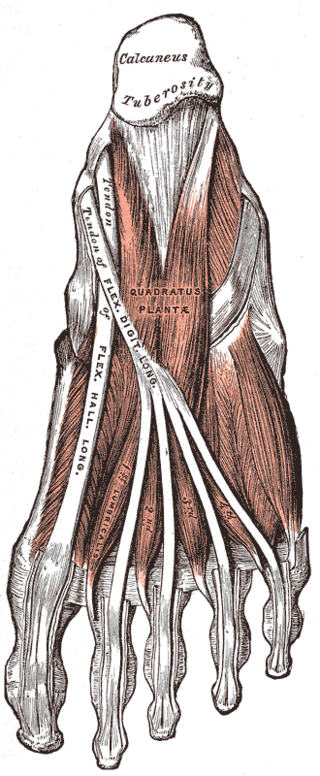
The quadratus plantae is separated from the muscles of the first layer by the lateral plantar vessels and nerve. It acts to aid in flexing the 2nd to 5th toes and is one of the few muscles in the foot with no homolog in the hand.

The lumbricals are four small skeletal muscles, accessory to the tendons of the flexor digitorum longus muscle. They are numbered from the medial side of the foot.
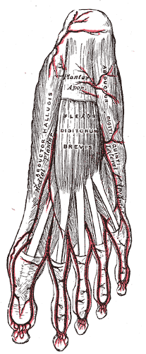
The abductor digiti minimi is a muscle which lies along the lateral (outer) border of the foot, and is in relation by its medial margin with the lateral plantar artery, vein and nerves.

In humans, the sole of the foot is anatomically referred to as the plantar aspect.

The medial plantar nerve is the larger of the two terminal divisions of the tibial nerve, which accompanies the medial plantar artery.

The interphalangeal joints of the foot are the joints between the phalanx bones of the toes in the feet.
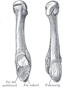
The fifth metatarsal bone is a long bone in the foot, and is palpable along the distal outer edges of the feet. It is the second smallest of the five metatarsal bones. The fifth metatarsal is analogous to the fifth metacarpal bone in the hand.

The extrinsic extensor muscles of the hand are located in the back of the forearm and have long tendons connecting them to bones in the hand, where they exert their action. Extrinsic denotes their location outside the hand. Extensor denotes their action which is to extend, or open flat, joints in the hand. They include the extensor carpi radialis longus (ECRL), extensor carpi radialis brevis (ECRB), extensor digitorum (ED), extensor digiti minimi (EDM), extensor carpi ulnaris (ECU), abductor pollicis longus (APL), extensor pollicis brevis (EPB), extensor pollicis longus (EPL), and extensor indicis (EI).


