Gastrocnemius muscle

Last updated
Gastrocnemius muscle
Sobo 1909 303 - Gastrocnemius muscle.png
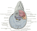
Right leg seen from behind.
Dissection video (2 min 44 s)
Details
Pronunciation /ˌɡæstrɒkˈnmiəs/ or /ˌɡæstrəkˈnmiəs/
Origin Proximal to articular surfaces of lateral condyle of femur and medial condyle of femur
Insertion Tendo calcaneus (Achilles tendon) into mid-posterior calcaneus
Artery Sural arteries
Nerve Tibial nerve from the sciatic, specifically, nerve roots S1–S2
Actions Plantar flexes foot, flexes knee
Antagonist Tibialis anterior muscle
Identifiers
TA98 A04.7.02.044
TA2 2657
FMA 22541
Anatomical terms of muscle

The gastrocnemius muscle (plural gastrocnemii) is a superficial two-headed muscle that is in the back part of the lower leg of humans. It is located superficial to the soleus in the posterior (back) compartment of the leg. It runs from its two heads just above the knee to the heel, extending across a total of three joints (knee, ankle and subtalar joints).

Contents

The muscle is named via Latin, from Greek γαστήρ (gaster) 'belly' or 'stomach' and κνήμη (knḗmē) 'leg', meaning 'stomach of the leg' (referring to the bulging shape of the calf).

Structure

Origin/proximal attachment

The lateral head originates from the lateral condyle of the femur, while the medial head originates from the medial condyle of the femur.

Insertion/distal attachment

Its other end forms a common tendon with the soleus muscle; this tendon is known as the calcaneal tendon or Achilles tendon and inserts onto the posterior surface of the calcaneus, or heel bone.

Relations

The gastrocnemius is located with the soleus in the posterior compartment of leg. It is considered a superficial muscle as it is located directly under skin, and its shape may often be visualized through the skin.

Beneath the gastrocnemius (farther from the skin) is the soleus muscle. Some anatomists consider both to be a single muscle—the triceps surae or "three-headed [muscle] of the calf"—since they share a common insertion via the Achilles tendon. The plantaris muscle and a portion of its tendon run between the two muscles, which is involved in "locking" the knee from the standing position. Since the anterior compartment of the leg is lateral to the tibia, the bulge of muscle medial to the tibia on the anterior side is actually the posterior compartment. The soleus is superficial to the mid-shaft of the tibia.

Variation

39% of individuals have a sesamoid bone called the fabella in the lateral (outer) head of the gastrocnemius muscle.[ citation needed ]

Function

Along with the soleus muscle, the gastrocnemius forms half of the calf muscle. Its function is plantar flexing the foot at the ankle joint and flexing the leg at the knee joint.

The gastrocnemius is primarily involved in running, jumping and other "fast" movements of leg, and to a lesser degree in walking and standing. This specialization is connected to the predominance of white muscle fibers (type II fast twitch) present in the gastrocnemius, as opposed to the soleus, which has more red muscle fibers (type I slow twitch) and is the primary active muscle when standing still. [1] [2]

Motor pathway

The plan to use the gastrocnemius in running, jumping, knee and plantar flexing is created in the precentral gyrus in the cerebrum of the brain. [3] Once a plan is produced, the signal is sent to and down an upper motor neuron. The signal is passed through the internal capsule and decussates, or crosses, in the medulla oblongata, specifically in the lateral corticospinal tract. [4] The signal continues down through the anterior horn of the spinal cord where the upper motor neuron synapses with the lower motor neuron. Signal propagation continues down the anterior rami (Lumbar 4-5 and Sacral 1-5) of the sacral plexus. The sciatic nerve branches off of the sacral plexus in which the tibial and common fibular nerves are wrapped in one sheath. The tibial nerve eventually separates from the sciatic nerve and innervates the gastrocnemius muscle.

Clinical significance

The gastrocnemius muscle is prone to spasms, which are painful, involuntary contractions of the muscle that may last several minutes. [5]

A severe ankle dorsiflexion force may result in a Medial Gastrocnemius Strain (MGS) injury of the muscle, commonly referred to as a "torn" or "strained" calf muscle, which is acutely painful and disabling. [6]

The gastrocnemius muscle may also become inflamed due to overuse. Anti-inflammatory medications and physical therapy (heat, massage, and stretching) may be useful.

Anatomical abnormalities involving the medial head of gastrocnemius muscle result in popliteal artery entrapment syndrome.

History

In a 1967 EMG study, Herman and Bragin concluded that its most important role was plantar flexing in large contractions and in rapid development of tension. [2]

Additional images

See also

Related Research Articles

<span class="mw-page-title-main">Foot</span> Anatomical structure found in vertebrates

The foot is an anatomical structure found in many vertebrates. It is the terminal portion of a limb which bears weight and allows locomotion. In many animals with feet, the foot is a separate organ at the terminal part of the leg made up of one or more segments or bones, generally including claws and/or nails.

<span class="mw-page-title-main">Human leg</span> Lower extremity or limb of the human body (foot, lower leg, thigh and hip)

The leg is the entire lower limb of the human body, including the foot, thigh or sometimes even the hip or buttock region. The major bones of the leg are the femur, tibia, and adjacent fibula. The thigh is between the hip and knee, while the calf (rear) and shin (front) are between the knee and foot.

<span class="mw-page-title-main">Knee</span> Leg joint in primates

In humans and other primates, the knee joins the thigh with the leg and consists of two joints: one between the femur and tibia, and one between the femur and patella. It is the largest joint in the human body. The knee is a modified hinge joint, which permits flexion and extension as well as slight internal and external rotation. The knee is vulnerable to injury and to the development of osteoarthritis.

<span class="mw-page-title-main">Sciatic nerve</span> Large nerve in humans and other animals

The sciatic nerve, also called the ischiadic nerve, is a large nerve in humans and other vertebrate animals which is the largest branch of the sacral plexus and runs alongside the hip joint and down the lower limb. It is the longest and widest single nerve in the human body, going from the top of the leg to the foot on the posterior aspect. The sciatic nerve has no cutaneous branches for the thigh. This nerve provides the connection to the nervous system for the skin of the lateral leg and the whole foot, the muscles of the back of the thigh, and those of the leg and foot. It is derived from spinal nerves L4 to S3. It contains fibers from both the anterior and posterior divisions of the lumbosacral plexus.

<span class="mw-page-title-main">Fibularis longus</span> Superficial muscle in the lateral compartment of the leg

In human anatomy, the fibularis longus is a superficial muscle in the lateral compartment of the leg. It acts to tilt the sole of the foot away from the midline of the body (eversion) and to extend the foot downward away from the body at the ankle.

<span class="mw-page-title-main">Tibia</span> Leg bone in vertebrates

The tibia, also known as the shinbone or shankbone, is the larger, stronger, and anterior (frontal) of the two bones in the leg below the knee in vertebrates ; it connects the knee with the ankle. The tibia is found on the medial side of the leg next to the fibula and closer to the median plane. The tibia is connected to the fibula by the interosseous membrane of leg, forming a type of fibrous joint called a syndesmosis with very little movement. The tibia is named for the flute tibia. It is the second largest bone in the human body, after the femur. The leg bones are the strongest long bones as they support the rest of the body.

<span class="mw-page-title-main">Fibula</span> Leg bone in vertebrates

The fibula or calf bone is a leg bone on the lateral side of the tibia, to which it is connected above and below. It is the smaller of the two bones and, in proportion to its length, the most slender of all the long bones. Its upper extremity is small, placed toward the back of the head of the tibia, below the knee joint and excluded from the formation of this joint. Its lower extremity inclines a little forward, so as to be on a plane anterior to that of the upper end; it projects below the tibia and forms the lateral part of the ankle joint.

<span class="mw-page-title-main">Popliteal artery</span> Continuation of the femoral artery that supplies the lower leg

The popliteal artery is a deeply placed continuation of the femoral artery opening in the distal portion of the adductor magnus muscle. It courses through the popliteal fossa and ends at the lower border of the popliteus muscle, where it branches into the anterior and posterior tibial arteries.

<span class="mw-page-title-main">Soleus muscle</span> Powerful muscle in the back part of the lower leg

In humans and some other mammals, the soleus is a powerful muscle in the back part of the lower leg. It runs from just below the knee to the heel and is involved in standing and walking. It is closely connected to the gastrocnemius muscle, and some anatomists consider this combination to be a single muscle, the triceps surae. Its name is derived from the Latin word "solea", meaning "sandal".

<span class="mw-page-title-main">Triceps surae muscle</span> Pair of muscles: gastrocnemius and the soleus

The triceps surae consists of two muscles located at the calf – the two-headed gastrocnemius and the soleus. These muscles both insert into the calcaneus, the bone of the heel of the human foot, and form the major part of the muscle of the posterior leg, commonly known as the calf muscle.

<span class="mw-page-title-main">Tibial nerve</span> Branch of the sciatic nerve

The tibial nerve is a branch of the sciatic nerve. The tibial nerve passes through the popliteal fossa to pass below the arch of soleus.

The biceps femoris is a muscle of the thigh located to the posterior, or back. As its name implies, it consists of two heads; the long head is considered part of the hamstring muscle group, while the short head is sometimes excluded from this characterization, as it only causes knee flexion and is activated by a separate nerve.

The semimembranosus muscle is the most medial of the three hamstring muscles in the thigh. It is so named because it has a flat tendon of origin. It lies posteromedially in the thigh, deep to the semitendinosus muscle. It extends the hip joint and flexes the knee joint.

<span class="mw-page-title-main">Semitendinosus muscle</span> One of the hamstring muscles; posterior part of the thigh

The semitendinosus is a long superficial muscle in the back of the thigh. It is so named because it has a very long tendon of insertion. It lies posteromedially in the thigh, superficial to the semimembranosus.

<span class="mw-page-title-main">Flexor digitorum longus muscle</span> Muscle located on the tibial side of the leg

The flexor digitorum longus muscle is situated on the tibial side of the leg. At its origin it is thin and pointed, but it gradually increases in size as it descends. It serves to flex the second, third, fourth, and fifth toes.

<span class="mw-page-title-main">Popliteus muscle</span> Muscle responsible for unlocking the knees during walking

The popliteus muscle in the leg is used for unlocking the knees when walking, by laterally rotating the femur on the tibia during the closed chain portion of the gait cycle. In open chain movements, the popliteus muscle medially rotates the tibia on the femur. It is also used when sitting down and standing up. It is the only muscle in the posterior (back) compartment of the lower leg that acts just on the knee and not on the ankle. The gastrocnemius muscle acts on both joints.

<span class="mw-page-title-main">Plantaris muscle</span> One of the superficial muscles of the superficial posterior compartment of the leg

The plantaris is one of the superficial muscles of the superficial posterior compartment of the leg, one of the fascial compartments of the leg.

<span class="mw-page-title-main">Sole (foot)</span> Bottom part of foot

In humans, the sole of the foot is anatomically referred to as the plantar aspect.

<span class="mw-page-title-main">Outline of human anatomy</span> Overview of and topical guide to human anatomy

The following outline is provided as an overview of and topical guide to human anatomy:

References

  1. Moore, Keith L.; Dalley, Arthur F.; Agur, Anne M. R. (2009). Clinically Oriented Anatomy. pp. 598–600. ISBN   978-1-60547-652-0.
  2. 1 2 Hamilton, Nancy; Luttgens, Kathryn (2001). Kinesiology: Scientific Basis of Human Motion (10th ed.). McGraw-Hill. ISBN   978-0-07-248910-1.
  3. Freberg, Laura (2015-01-01). Discovering Behavioral Neuroscience: An Introduction to Biological Psychology. Cengage Learning. ISBN   9781305687738.
  4. Saladin, Kenneth (2015). Anatomy & Physiology: The Unity of Form and Function. McGraw-Hill Education.
  5. "Nighttime Leg Cramps". WebMD. August 19, 2010. Retrieved March 7, 2012.
  6. "Calf Muscle Tear". physioworks.com.au. Retrieved 2020-02-09.