Popliteus muscle

Last updated
Popliteus muscle
Posterior compartment of leg - popliteus muscle.png
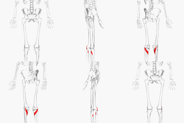
Popliteus muscle (shown in red)
Dissection video (2 min 22 s)
Details
Origin Lateral femoral epicondyle
Insertion Posterior surface of the tibia proximal to the soleus line
Artery Popliteal artery
Nerve Tibial nerve
Actions Medially rotates tibia on the femur if the femur is fixed (sitting down) or laterally rotates femur on the tibia if tibia is fixed (standing up), unlocks the knee to allow flexion (bending), helps to prevent the forward dislocation of the femur while crouching
Identifiers
Latin musculus popliteus, poplit=ham (pit) of the knee
TA98 A04.7.02.050
TA2 2665
FMA 22590
Anatomical terms of muscle

The popliteus muscle in the leg is used for unlocking the knees when walking, by laterally rotating the femur on the tibia during the closed chain portion of the gait cycle (one with the foot in contact with the ground). In open chain movements (when the involved limb is not in contact with the ground), the popliteus muscle medially rotates the tibia on the femur. It is also used when sitting down and standing up. It is the only muscle in the posterior (back) compartment of the lower leg that acts just on the knee and not on the ankle. The gastrocnemius muscle acts on both joints.
Contents

Structure

The popliteus muscle originates from the lateral surface of the lateral condyle of the femur by a rounded tendon. [1] Its fibers pass downward and medially. It inserts onto the posterior surface of tibia, above the soleal line. [1] The popliteus tendon runs beneath the lateral collateral ligament and tendon of biceps femoris. The muscle also runs above the lateral meniscus but has no connection with the meniscus in 45% of the cases, but has strong connection with it in 17.5% of the cases. Therefore, popliteus muscle is extrasynovial, extra-articular, and intracapsular. [2]

Nerve supply

The popliteus muscle is supplied by the tibial nerve, from spinal roots L5 and S1.

Variation

There is sometimes an additional head from the sesamoid bone in the lateral (outer) head of the gastrocnemius muscle.

Rarely an additional inconstant muscle; the popliteus minor is seen. It originates from the femur on the inner side of the plantaris muscle and inserts into the posterior ligament of the knee-joint.

Peroneotibialis, 14% of population. Origin is inner side of the head of the fibula, insertion into the upper end of the oblique line of the tibia, it lies beneath the popliteus. [3]

Another variant, the cyamella, is a small sesamoid bone embedded in the tendon of the popliteus muscle. It is rarely seen in humans, with prevalence rates from 0.57–1.8%, [4] but has been described more often in other primates and certain other animals. [5]

Function

The popliteus assists in flexing the leg upon the thigh; when the leg is flexed, it will rotate the tibia inward.

It is especially called into action at the beginning of the act of bending the knee, in as much as it produces the slight inward rotation of the tibia, which is essential in the early stage of this movement. [6]

When the knee is in full extension, the femur slightly medially rotates on the tibia to lock the knee joint in place. Popliteus is often referred to as the "Key" to unlocking the knee since it begins knee flexion by laterally rotating the femur on the tibia. [6]

Popliteus is also attached to the lateral meniscus in the knee and draws it posteriorly during knee flexion to prevent crushing the meniscus between the tibia and femur as the knee flexes.

Additional images

See also

Surgery

Injury to the Popliteus causes posterolateral rotatory instability of knee. This can be treated with Arthroscopic Popliteus Sling reconstruction using the popliteus portal. [7]

Related Research Articles

<span class="mw-page-title-main">Foot</span> Anatomical structure found in vertebrates

The foot is an anatomical structure found in many vertebrates. It is the terminal portion of a limb which bears weight and allows locomotion. In many animals with feet, the foot is a separate organ at the terminal part of the leg made up of one or more segments or bones, generally including claws and/or nails.

<span class="mw-page-title-main">Human leg</span> Lower extremity or limb of the human body (foot, lower leg, thigh and hip)

The leg is the entire lower limb of the human body, including the foot, thigh or sometimes even the hip or buttock region. The major bones of the leg are the femur, tibia, and adjacent fibula. The thigh is between the hip and knee, while the calf (rear) and shin (front) are between the knee and foot.

<span class="mw-page-title-main">Knee</span> Leg joint in primates

In humans and other primates, the knee joins the thigh with the leg and consists of two joints: one between the femur and tibia, and one between the femur and patella. It is the largest joint in the human body. The knee is a modified hinge joint, which permits flexion and extension as well as slight internal and external rotation. The knee is vulnerable to injury and to the development of osteoarthritis.

<span class="mw-page-title-main">Hamstring</span> Any of the three tendons in the thigh

In human anatomy, a hamstring is any one of the three posterior thigh muscles between the hip and the knee.

<span class="mw-page-title-main">Tibia</span> Leg bone in vertebrates

The tibia, also known as the shinbone or shankbone, is the larger, stronger, and anterior (frontal) of the two bones in the leg below the knee in vertebrates ; it connects the knee with the ankle. The tibia is found on the medial side of the leg next to the fibula and closer to the median plane. The tibia is connected to the fibula by the interosseous membrane of leg, forming a type of fibrous joint called a syndesmosis with very little movement. The tibia is named for the flute tibia. It is the second largest bone in the human body, after the femur. The leg bones are the strongest long bones as they support the rest of the body.

<span class="mw-page-title-main">Posterior cruciate ligament</span> One of four major ligaments of the knee

The posterior cruciate ligament (PCL) is a ligament in each knee of humans and various other animals. It works as a counterpart to the anterior cruciate ligament (ACL). It connects the posterior intercondylar area of the tibia to the medial condyle of the femur. This configuration allows the PCL to resist forces pushing the tibia posteriorly relative to the femur.

<span class="mw-page-title-main">Anterior cruciate ligament</span> Type of cruciate ligament in the human knee

The anterior cruciate ligament (ACL) is one of a pair of cruciate ligaments in the human knee. The two ligaments are also called "cruciform" ligaments, as they are arranged in a crossed formation. In the quadruped stifle joint, based on its anatomical position, it is also referred to as the cranial cruciate ligament. The term cruciate translates to cross. This name is fitting because the ACL crosses the posterior cruciate ligament to form an "X". It is composed of strong, fibrous material and assists in controlling excessive motion. This is done by limiting mobility of the joint. The anterior cruciate ligament is one of the four main ligaments of the knee, providing 85% of the restraining force to anterior tibial displacement at 30 and 90° of knee flexion. The ACL is the most injured ligament of the four located in the knee.

<span class="mw-page-title-main">Popliteal artery</span> Continuation of the femoral artery that supplies the lower leg

The popliteal artery is a deeply placed continuation of the femoral artery opening in the distal portion of the adductor magnus muscle. It courses through the popliteal fossa and ends at the lower border of the popliteus muscle, where it branches into the anterior and posterior tibial arteries.

<span class="mw-page-title-main">Soleus muscle</span> Powerful muscle in the back part of the lower leg

In humans and some other mammals, the soleus is a powerful muscle in the back part of the lower leg. It runs from just below the knee to the heel and is involved in standing and walking. It is closely connected to the gastrocnemius muscle, and some anatomists consider this combination to be a single muscle, the triceps surae. Its name is derived from the Latin word "solea", meaning "sandal".

<span class="mw-page-title-main">Gastrocnemius muscle</span> Calf muscle

The gastrocnemius muscle is a superficial two-headed muscle that is in the back part of the lower leg of humans. It is located superficial to the soleus in the posterior (back) compartment of the leg. It runs from its two heads just above the knee to the heel, extending across a total of three joints.

<span class="mw-page-title-main">Tibial nerve</span> Branch of the sciatic nerve

The tibial nerve is a branch of the sciatic nerve. The tibial nerve passes through the popliteal fossa to pass below the arch of soleus.

The biceps femoris is a muscle of the thigh located to the posterior, or back. As its name implies, it consists of two heads; the long head is considered part of the hamstring muscle group, while the short head is sometimes excluded from this characterization, as it only causes knee flexion and is activated by a separate nerve.

The semimembranosus muscle is the most medial of the three hamstring muscles in the thigh. It is so named because it has a flat tendon of origin. It lies posteromedially in the thigh, deep to the semitendinosus muscle. It extends the hip joint and flexes the knee joint.

<span class="mw-page-title-main">Plantaris muscle</span> One of the superficial muscles of the superficial posterior compartment of the leg

The plantaris is one of the superficial muscles of the superficial posterior compartment of the leg, one of the fascial compartments of the leg.

<span class="mw-page-title-main">Stifle joint</span>

The stifle joint is a complex joint in the hind limbs of quadruped mammals such as the sheep, horse or dog. It is the equivalent of the human knee and is often the largest synovial joint in the animal's body. The stifle joint joins three bones: the femur, patella, and tibia. The joint consists of three smaller ones: the femoropatellar joint, medial femorotibial joint, and lateral femorotibial joint.

<span class="mw-page-title-main">Unhappy triad</span> Medical condition of the knee

The unhappy triad, also known as a blown knee among other names, is an injury to the anterior cruciate ligament, medial collateral ligament, and meniscus. Analysis during the 1990s indicated that this 'classic' O'Donoghue triad is actually an unusual clinical entity among athletes with knee injuries. Some authors mistakenly believe that in this type of injury, "combined anterior cruciate and medial collateral ligament disruptions that were incurred during athletic endeavors" always present with concomitant medial meniscus injury. However, the 1990 analysis showed that lateral meniscus tears are more common than medial meniscus tears in conjunction with sprains of the ACL.

<span class="mw-page-title-main">Articular capsule of the knee joint</span>

The articular capsule of the knee joint is the wide and lax joint capsule of the knee. It is thin in front and at the side, and contains the patella, ligaments, menisci, and bursae of the knee. The capsule consists of an inner synovial membrane, and an outer fibrous membrane separated by fatty deposits anteriorly and posteriorly.

Posterolateral corner injuries of the knee are injuries to a complex area formed by the interaction of multiple structures. Injuries to the posterolateral corner can be debilitating to the person and require recognition and treatment to avoid long term consequences. Injuries to the PLC often occur in combination with other ligamentous injuries to the knee; most commonly the anterior cruciate ligament (ACL) and posterior cruciate ligament (PCL). As with any injury, an understanding of the anatomy and functional interactions of the posterolateral corner is important to diagnosing and treating the injury.

<span class="mw-page-title-main">Medial knee injuries</span> Medical condition

Medial knee injuries are the most common type of knee injury. The medial ligament complex of the knee consists of:

<span class="mw-page-title-main">Posterior cruciate ligament injury</span> Medical condition

The function of the posterior cruciate ligament (PCL) is to prevent the femur from sliding off the anterior edge of the tibia and to prevent the tibia from displacing posterior to the femur. Common causes of PCL injuries are direct blows to the flexed knee, such as the knee hitting the dashboard in a car accident or falling hard on the knee, both instances displacing the tibia posterior to the femur.

References

PD-icon.svgThis article incorporates text in the public domain from page 484 of the 20th edition of Gray's Anatomy (1918)

  1. 1 2 Strickland, Justin P.; Fester, Eric W.; Noyes, Frank R. (2017-01-01), Noyes, Frank R.; Barber-Westin, Sue D. (eds.), "2 - Lateral and Posterior Knee Anatomy", Noyes' Knee Disorders: Surgery, Rehabilitation, Clinical Outcomes (Second Edition), Elsevier, pp. 23–35, doi:10.1016/b978-0-323-32903-3.00002-0, ISBN   978-0-323-32903-3 , retrieved 2021-03-01
  2. Jadhav, Siddharth P.; More, Snehal R.; Riascos, Roy F.; Lemos, Diego F.; Swischuk, Leonard E. (March 2014). "Comprehensive Review of the Anatomy, Function, and Imaging of the Popliteus and Associated Pathologic Conditions". RadioGraphics. 34 (2): 496–513. doi:10.1148/rg.342125082. ISSN   0271-5333. PMID   24617694.
  3. Gray, Henry. 1918. Anatomy of the Human Body. Page 485
  4. Berthaume, Michael A.; Bull, Anthony M. J. (July 2021). "Cyamella (a popliteal sesamoid bone) prevalence: A systematic review, meta‐analysis, and proposed classification system". Clinical Anatomy. 34 (5): 810–820. doi:10.1002/ca.23743. hdl: 10044/1/88423 . ISSN   0897-3806. PMID   33905585. S2CID   233427472.
  5. Akansel, Gur; Inan, Nagihan; Sarisoy, H. Tahsin; Anik, Yonca; Akansel, Sertaç (2006). "Popliteus muscle sesamoid bone (Cyamella): Appearance on radiographs, CT and MRI". Surgical and Radiologic Anatomy. 28 (6): 642–645. doi:10.1007/s00276-006-0134-8. PMID   17066262. S2CID   13339926.
  6. 1 2 Wood, Addison; Boren, Morgan; Dodgen, Taylor; Wagner, Russell; Patterson, Rita M. (March 2020). "Muscular architecture of the popliteus muscle and the basic science implications". The Knee. 27 (2): 308–314. doi:10.1016/j.knee.2019.12.001. ISSN   1873-5800. PMID   31954610. S2CID   210831808.
  7. Kodkani, Pranjal (1 March 2011). "Arthroscopic Popliteus Sling Reconstruction—The Popliteus Portal". Techniques in Knee Surgery. 10 (2011): 58–64. doi:10.1097/BTK.0B013E31820D6C7E. S2CID   76366926.