Lateral meniscus

Last updated
Lateral meniscus
Blausen 0597 KneeAnatomy Side.png
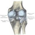
Knee from the side, with lateral meniscus simply labeled as "meniscus".
Gray349.png
Head of right tibia seen from above, showing menisci and attachments of ligaments.
Details
Identifiers
Latin meniscus lateralis
TA98 A03.6.08.002
TA2 1885
FMA 44631
Anatomical terminology

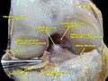
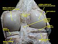
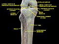
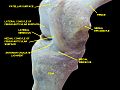
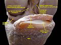
The lateral meniscus (external semilunar fibrocartilage) is a fibrocartilaginous band that spans the lateral side of the interior of the knee joint. It is one of two menisci of the knee, the other being the medial meniscus. It is nearly circular and covers a larger portion of the articular surface than the medial. It can occasionally be injured or torn by twisting the knee or applying direct force, as seen in contact sports.

Contents

Structure

The lateral meniscus is grooved laterally for the tendon of the popliteus, which separates it from the fibular collateral ligament.

Its anterior end is attached in front of the intercondyloid eminence of the tibia, lateral to, and behind, the anterior cruciate ligament, with which it blends; the posterior end is attached behind the intercondyloid eminence of the tibia and in front of the posterior end of the medial meniscus.

The anterior attachment of the lateral meniscus is twisted on itself so that its free margin looks backward and upward, its anterior end resting on a sloping shelf of bone on the front of the lateral process of the intercondyloid eminence.

Close to its posterior attachment it sends off a strong fasciculus, the ligament of Wrisberg, which passes upward and medialward, to be inserted into the medial condyle of the femur, immediately behind the attachment of the posterior cruciate ligament.

The lateral meniscus gives off from its anterior convex margin a fasciculus which forms the transverse ligament.

Variation

Occasionally a small fasciculus passes forward to be inserted into the lateral part of the anterior cruciate ligament.

Clinical significance

The lateral meniscus is less likely to be injured or torn than the medial meniscus. Diagnosis of lateral meniscus tear is done with McMurray's test. If a tear is detected, treatment depends on the type and size of the tear. Small tears can be treated conservatively, with rest, ice, and pain medications until the pain is under control, then exercise may be started with gradually increasing intensity, to improve range of motion and decrease swelling. More severe tears of the lateral meniscus require surgical repair or removal, which can often be done arthroscopically. Swelling and stiffness of the knee can occur when you have a torn lateral meniscus.

Additional images

See also

Related Research Articles

<span class="mw-page-title-main">Femur</span> Thigh bone

The femur, or thigh bone is the only bone in the thigh. The thigh is the region of the lower limb between the hip and the knee. In many four-legged animals the femur is the upper bone of the hindleg.

<span class="mw-page-title-main">Knee</span> Leg joint in primates

In humans and other primates, the knee joins the thigh with the leg and consists of two joints: one between the femur and tibia, and one between the femur and patella. It is the largest joint in the human body. The knee is a modified hinge joint, which permits flexion and extension as well as slight internal and external rotation. The knee is vulnerable to injury and to the development of osteoarthritis.

<span class="mw-page-title-main">Humerus</span> Long bone of the upper arm

The humerus is a long bone in the arm that runs from the shoulder to the elbow. It connects the scapula and the two bones of the lower arm, the radius and ulna, and consists of three sections. The humeral upper extremity consists of a rounded head, a narrow neck, and two short processes. The body is cylindrical in its upper portion, and more prismatic below. The lower extremity consists of 2 epicondyles, 2 processes, and 3 fossae. As well as its true anatomical neck, the constriction below the greater and lesser tubercles of the humerus is referred to as its surgical neck due to its tendency to fracture, thus often becoming the focus of surgeons.

<span class="mw-page-title-main">Tibia</span> Leg bone in vertebrates

The tibia, also known as the shinbone or shankbone, is the larger, stronger, and anterior (frontal) of the two bones in the leg below the knee in vertebrates ; it connects the knee with the ankle. The tibia is found on the medial side of the leg next to the fibula and closer to the median plane. The tibia is connected to the fibula by the interosseous membrane of leg, forming a type of fibrous joint called a syndesmosis with very little movement. The tibia is named for the flute tibia. It is the second largest bone in the human body, after the femur. The leg bones are the strongest long bones as they support the rest of the body.

<span class="mw-page-title-main">Fibula</span> Leg bone in vertebrates

The fibula or calf bone is a leg bone on the lateral side of the tibia, to which it is connected above and below. It is the smaller of the two bones and, in proportion to its length, the most slender of all the long bones. Its upper extremity is small, placed toward the back of the head of the tibia, below the knee joint and excluded from the formation of this joint. Its lower extremity inclines a little forward, so as to be on a plane anterior to that of the upper end; it projects below the tibia and forms the lateral part of the ankle joint.

<span class="mw-page-title-main">Posterior cruciate ligament</span> One of four major ligaments of the knee

The posterior cruciate ligament (PCL) is a ligament in each knee of humans and various other animals. It works as a counterpart to the anterior cruciate ligament (ACL). It connects the posterior intercondylar area of the tibia to the medial condyle of the femur. This configuration allows the PCL to resist forces pushing the tibia posteriorly relative to the femur.

<span class="mw-page-title-main">Anterior cruciate ligament</span> Type of cruciate ligament in the human knee

The anterior cruciate ligament (ACL) is one of a pair of cruciate ligaments in the human knee. The two ligaments are also called "cruciform" ligaments, as they are arranged in a crossed formation. In the quadruped stifle joint, based on its anatomical position, it is also referred to as the cranial cruciate ligament. The term cruciate translates to cross. This name is fitting because the ACL crosses the posterior cruciate ligament to form an "X". It is composed of strong, fibrous material and assists in controlling excessive motion. This is done by limiting mobility of the joint. The anterior cruciate ligament is one of the four main ligaments of the knee, providing 85% of the restraining force to anterior tibial displacement at 30 and 90° of knee flexion. The ACL is the most injured ligament of the four located in the knee.

<span class="mw-page-title-main">Medial meniscus</span> Cartilage formation in the human knee

The medial meniscus is a fibrocartilage semicircular band that spans the knee joint medially, located between the medial condyle of the femur and the medial condyle of the tibia. It is also referred to as the internal semilunar fibrocartilage. The medial meniscus has more of a crescent shape while the lateral meniscus is more circular. The anterior aspects of both menisci are connected by the transverse ligament. It is a common site of injury, especially if the knee is twisted.

The knee examination, in medicine and physiotherapy, is performed as part of a physical examination, or when a patient presents with knee pain or a history that suggests a pathology of the knee joint.

<span class="mw-page-title-main">Lower extremity of femur</span>

The lower extremity of femur is the lower end of the femur in human and other animals, closer to the knee. It is larger than the upper extremity of femur, is somewhat cuboid in form, but its transverse diameter is greater than its antero-posterior; it consists of two oblong eminences known as the lateral condyle and medial condyle.

<span class="mw-page-title-main">Fibular collateral ligament</span> Collateral fibular ligament

The lateral collateral ligament is an extrinsic ligament of the knee located on the lateral side of the knee. Its superior attachment is at the lateral epicondyle of the femur ; its inferior attachment is at the lateral aspect of the head of fibula. The LCL is not fused with the joint capsule. Inferiorly, the LCL splits the tendon of insertion of the biceps femoris muscle.

<span class="mw-page-title-main">Patellar tendon</span> Tendon in the human knee

The patellar tendon is the distal portion of the common tendon of the quadriceps femoris, which is continued from the patella to the tibial tuberosity. It is also sometimes called the patellar ligament as it forms a bone to bone connection when the patella is fully ossified.

<span class="mw-page-title-main">Unhappy triad</span> Medical condition of the knee

The unhappy triad, also known as a blown knee among other names, is an injury to the anterior cruciate ligament, medial collateral ligament, and meniscus. Analysis during the 1990s indicated that this 'classic' O'Donoghue triad is actually an unusual clinical entity among athletes with knee injuries. Some authors mistakenly believe that in this type of injury, "combined anterior cruciate and medial collateral ligament disruptions that were incurred during athletic endeavors" always present with concomitant medial meniscus injury. However, the 1990 analysis showed that lateral meniscus tears are more common than medial meniscus tears in conjunction with sprains of the ACL.

<span class="mw-page-title-main">Malleolus</span> Ankle bone protrusion

A malleolus is the bony prominence on each side of the human ankle.

<span class="mw-page-title-main">Articular capsule of the knee joint</span>

The articular capsule of the knee joint is the wide and lax joint capsule of the knee. It is thin in front and at the side, and contains the patella, ligaments, menisci, and bursae of the knee. The capsule consists of an inner synovial membrane, and an outer fibrous membrane separated by fatty deposits anteriorly and posteriorly.

<span class="mw-page-title-main">Coronary ligament of the knee</span>

The coronary ligaments of the knee are portions of the joint capsule which connect the inferior edges of the fibrocartilaginous menisci to the periphery of the tibial plateaus.

<span class="mw-page-title-main">Transverse ligament of knee</span>

The transverse or [anterior] meniscomeniscal ligament is a ligament in the knee joint that connects the anterior convex margin of the lateral meniscus to the anterior end of the medial meniscus.

Posterolateral corner injuries of the knee are injuries to a complex area formed by the interaction of multiple structures. Injuries to the posterolateral corner can be debilitating to the person and require recognition and treatment to avoid long term consequences. Injuries to the PLC often occur in combination with other ligamentous injuries to the knee; most commonly the anterior cruciate ligament (ACL) and posterior cruciate ligament (PCL). As with any injury, an understanding of the anatomy and functional interactions of the posterolateral corner is important to diagnosing and treating the injury.

<span class="mw-page-title-main">Medial knee injuries</span> Medical condition

Medial knee injuries are the most common type of knee injury. The medial ligament complex of the knee consists of:

<span class="mw-page-title-main">Intercondylar area</span>

The intercondylar area is the separation between the medial and lateral condyle on the upper extremity of the tibia. The anterior and posterior cruciate ligaments and the menisci attach to the intercondylar area.

References

PD-icon.svgThis article incorporates text in the public domain from page 343 of the 20th edition of Gray's Anatomy (1918)