
The tibia, also known as the shinbone or shankbone, is the larger, stronger, and anterior (frontal) of the two bones in the leg below the knee in vertebrates, and it connects the knee with the ankle bones. The tibia is found on the medial side of the leg next to the fibula and closer to the median plane or centre-line. The tibia is connected to the fibula by the interosseous membrane of the leg, forming a type of fibrous joint called a syndesmosis with very little movement. The tibia is named for the flute tibia. It is the second largest bone in the human body next to the femur. The leg bones are the strongest long bones as they support the rest of the body.
The ankle, or the talocrural region, is the region where the foot and the leg meet. The ankle includes three joints: the ankle joint proper or talocrural joint, the subtalar joint, and the inferior tibiofibular joint. The movements produced at this joint are dorsiflexion and plantarflexion of the foot. In common usage, the term ankle refers exclusively to the ankle region. In medical terminology, "ankle" can refer broadly to the region or specifically to the talocrural joint.
Pott's fracture, also known as Pott's syndrome I and Dupuytren fracture, is an archaic term loosely applied to a variety of bimalleolar ankle fractures. The injury is caused by a combined abduction external rotation from an eversion force. This action strains the sturdy medial (deltoid) ligament of the ankle, often tearing off the medial malleolus due to its strong attachment. The talus then moves laterally, shearing off the lateral malleolus or, more commonly, breaking the fibula superior to the tibiofibular syndesmosis. If the tibia is carried anteriorly, the posterior margin of the distal end of the tibia is also sheared off by the talus. A fractured fibula in addition to detaching the medial malleolus will tear the tibiofibular syndesmosis. The combined fracture of the medial malleolus, lateral malleolus, and the posterior margin of the distal end of the tibia is known as a "trimalleolar fracture".

The tibialis anterior is a muscle in humans that originates in the upper two-thirds of the lateral (outside) surface of the tibia and inserts into the medial cuneiform and first metatarsal bones of the foot. It acts to dorsiflex and invert the foot. This muscle is mostly located near the shin.

In human anatomy, the subtalar joint, also known as the talocalcaneal joint, is a joint of the foot. It occurs at the meeting point of the talus and the calcaneus.
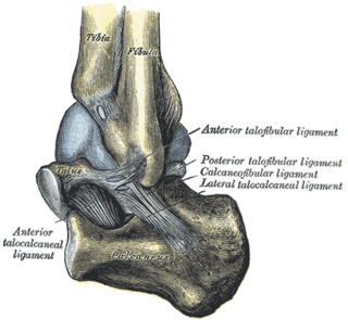
The anterior talocalcaneal ligament is a ligament in the foot.

The medial talocalcaneal ligament connects the medial tubercle of the back of the talus with the back of the sustentaculum tali.

In the sphenoid bone, the anterior boundary of the sella turcica is completed by two small eminences, one on either side, called the anterior clinoid processes, while the posterior boundary is formed by a square-shaped plate of bone, the dorsum sellæ, ending at its superior angles in two tubercles, the posterior clinoid processes, the size and form of which vary considerably in different individuals. The posterior clinoid processes deepen the sella turcica, and give attachment to the tentorium cerebelli.
The flexor retinaculum of foot is a strong fibrous band, extending from the bony ankle prominence (malleolus) above, to the margin of the heelbone (calcaneus) below, converting a series of bony grooves in this situation into canals for the passage of the tendons of the flexor muscles and the posterior tibial vessels and tibial nerve into the sole of the foot.

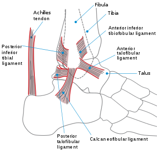
The anterior ligament of the lateral malleolus is a flat, triangular band of fibers, broader below than above, which extends obliquely downward and lateralward between the adjacent margins of the tibia and fibula, on the front aspect of the syndesmosis.
The inferior transverse ligament of the tibiofibular syndesmosis is a connective tissue structure in the lower leg that lies in front of the posterior ligament. It is a strong, thick band, of yellowish fibers which passes transversely across the back of the ankle joint, from the lateral malleolus to the posterior border of the articular surface of the tibia, almost as far as its malleolar process.
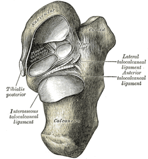
The talocalcaneonavicular joint is a ball and socket joint: the rounded head of the talus being received into the concavity formed by the posterior surface of the navicular, the anterior articular surface of the calcaneus, and the upper surface of the plantar calcaneonavicular ligament.
The plantar calcaneonavicular ligament is a complex of three ligaments on the underside of the foot that connect the calcaneus with the navicular bone.
The deltoid ligament is a strong, flat, triangular band, attached, above, to the apex and anterior and posterior borders of the medial malleolus. The deltoid ligament is composed of: 1. Anterior tibiotalar ligament 2. Tibiocalcaneal ligament 3. Posterior tibiotalar ligament 4. Tibionavicular ligament. It consists of two sets of fibers, superficial and deep.
The lateral collateral ligament of ankle joint are ligaments of the ankle which attach to the fibula.
The posterior talofibular ligament, runs almost horizontally from the malleolar fossa of the lateral malleolus of the fibula to a prominent tubercle on the posterior surface of the talus immediately lateral to the groove for the tendon of the flexor hallucis longus.
The anterior talofibular ligament is a ligament in the ankle. It passes from the anterior margin of the fibular malleolus, anteriorly and medially, to the talus bone, in front of its lateral articular facet. It is one of the lateral ligaments of the ankle and prevents the foot from sliding forward in relation to the shin. It is the most commonly injured ligament in a sprained ankle—from an inversion injury—and will allow a positive anterior drawer test of the ankle if completely torn.

A malleolus is the bony prominence on each side of the human ankle.
The public domain consists of all the creative works to which no exclusive intellectual property rights apply. Those rights may have expired, been forfeited, expressly waived, or may be inapplicable.

Gray's Anatomy is an English language textbook of human anatomy originally written by Henry Gray and illustrated by Henry Vandyke Carter. Earlier editions were called Anatomy: Descriptive and Surgical, Anatomy of the Human Body and Gray's Anatomy: Descriptive and Applied, but the book's name is commonly shortened to, and later editions are titled, Gray's Anatomy. The book is widely regarded as an extremely influential work on the subject, and has continued to be revised and republished from its initial publication in 1858 to the present day. The latest edition of the book, the 41st, was published in September 2015.
