Iliofemoral ligament

Last updated
Iliofemoral
Gray339.png
Right hip-joint from the front. (Iliofemoral ligament visible at center.)
Gray344.png
Structures surrounding right hip-joint. (Iliofemoral ligament labeled at upper left.)
Details
From ilium (anterior inferior iliac spine)
To femur (intertrochanteric line)
Identifiers
Latin ligamentum iliofemorale
TA98 A03.6.07.003
TA2 1875
FMA 42993
Anatomical terminology

The iliofemoral ligament is a thick and very tough triangular capsular ligament of the hip joint situated anterior to this joint. It attaches superiorly at the inferior portion of the anterior inferior iliac spine and adjacent portion of the margin of the acetabulum; it attaches inferiorly at the intertrochanteric line. [1]

Contents

It is also referred to as the Y-ligament (see below). the ligament of Bigelow , the ligament of Bertin and any combinations of these names.

With a force strength exceeding 350 kg (772 lbs), [2] the iliofemoral ligament is not only stronger than the two other ligaments of the hip joint, the ischiofemoral and the pubofemoral, but also the strongest ligament in the human body and as such is an important constraint to the hip joint. [3]

Structure

The ligament is triangular in shape, with its apex represented by its pelvic attachment. The ligament has two though outer bands; it is thinner and weaker centrally. [1] As the lateral portion is twisted like a screw, the two parts together take the form of an inverted Y. [4] [ verification needed ]

Arising from the anterior inferior iliac spine and the rim of the acetabulum, the iliofemoral ligament spreads obliquely downwards and laterally to the intertrochanteric line on the anterior side of the femoral head. It is divided into two parts or bands which act differently: the transverse part above, is strong and runs parallel to the axis of the femoral neck. The descending part below, is weaker and runs parallel to the femoral shaft. [4] [ verification needed ]

It is intimately connected with the joint capsule, and serves to strengthen the joint by resisting hyperextension. Its upper band is sometimes named the iliotrochanteric ligament. Between the two bands is a thinner part of the capsule. In some cases there is no division, and the ligament spreads out into a flat triangular band which is attached to the whole length of the intertrochanteric line.[ citation needed ]

Function

In a standing posture, when the pelvis is tilted posteriorly, the ligament is twisted and tense, which prevents the trunk from falling backwards and the posture is maintained without the need for muscular activity. In this position the ligament also keeps the femoral head pressed into the acetabulum. [4]

As the hip flexes, the tension in the ligament is reduced and the amount of possible rotations in the hip joint is increased, which permits the pelvis to tilt backwards into its sitting angle. Lateral rotation and adduction in the hip joint is controlled by the strong transversal part, while the descending part limits medial rotation. [4]

Turnout used in the classical ballet style requires a great deal of flexibility in this ligament. As does the front split where the rear leg is hyper-extended at the hip. Many externally rotate the rear leg while doing a front split, this external rotation when the hip is not flexed stretches the ligament even more. This "martial arts split" is distinguished by the rear knee pointing outward sideways (usually the foot along with it) rather than pointing straight down with the patella facing the floor, in a pure extension front split.

Additional images

Related Research Articles

<span class="mw-page-title-main">Human leg</span> Lower extremity or limb of the human body (foot, lower leg, thigh and hip)

The leg is the entire lower limb of the human body, including the foot, thigh or sometimes even the hip or buttock region. The major bones of the leg are the femur, tibia, and adjacent fibula. The thigh is between the hip and knee, while the calf (rear) and shin (front) are between the knee and foot.

<span class="mw-page-title-main">Hip</span> Anatomical region between the torso and the legs, holding the buttocks and genital region

In vertebrate anatomy, the hip, or coxa(pl.: coxae) in medical terminology, refers to either an anatomical region or a joint on the outer (lateral) side of the pelvis.

<span class="mw-page-title-main">Psoas major muscle</span> Long fusiform muscle located in the lumbar region

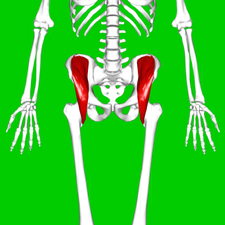
The psoas major is a long fusiform muscle located in the lateral lumbar region between the vertebral column and the brim of the lesser pelvis. It joins the iliacus muscle to form the iliopsoas. In animals, this muscle is equivalent to the tenderloin.

<span class="mw-page-title-main">Iliacus muscle</span> Flat, triangular muscle which fills the iliac fossa

The iliacus is a flat, triangular muscle which fills the iliac fossa. It forms the lateral portion of iliopsoas, providing flexion of the thigh and lower limb at the acetabulofemoral joint.

<span class="mw-page-title-main">Quadratus femoris muscle</span> One of six small hip muscles in the lateral rotator group

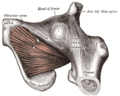
The quadratus femoris is a flat, quadrilateral skeletal muscle. Located on the posterior side of the hip joint, it is a strong external rotator and adductor of the thigh, but also acts to stabilize the femoral head in the acetabulum. The quadratus femoris is used in Meyer's muscle pedicle grafting to prevent avascular necrosis of femur head.

<span class="mw-page-title-main">Iliopsoas</span> Joined psoas and the iliacus muscles

The iliopsoas muscle refers to the joined psoas major and the iliacus muscles. The two muscles are separate in the abdomen, but usually merge in the thigh. They are usually given the common name iliopsoas. The iliopsoas muscle joins to the femur at the lesser trochanter. It acts as the strongest flexor of the hip.

<span class="mw-page-title-main">Lumbar plexus</span> Web of nerves in the lower spine

The lumbar plexus is a web of nerves in the lumbar region of the body which forms part of the larger lumbosacral plexus. It is formed by the divisions of the first four lumbar nerves (L1-L4) and from contributions of the subcostal nerve (T12), which is the last thoracic nerve. Additionally, the ventral rami of the fourth lumbar nerve pass communicating branches, the lumbosacral trunk, to the sacral plexus. The nerves of the lumbar plexus pass in front of the hip joint and mainly support the anterior part of the thigh.

<span class="mw-page-title-main">Sacrospinous ligament</span>

The sacrospinous ligament is a thin, triangular ligament in the human pelvis. The base of the ligament is attached to the outer edge of the sacrum and coccyx, and the tip of the ligament attaches to the spine of the ischium, a bony protuberance on the human pelvis. Its fibres are intermingled with the sacrotuberous ligament.

<span class="mw-page-title-main">Medial meniscus</span> Cartilage formation in the human knee

The medial meniscus is a fibrocartilage semicircular band that spans the knee joint medially, located between the medial condyle of the femur and the medial condyle of the tibia. It is also referred to as the internal semilunar fibrocartilage. The medial meniscus has more of a crescent shape while the lateral meniscus is more circular. The anterior aspects of both menisci are connected by the transverse ligament. It is a common site of injury, especially if the knee is twisted.

<span class="mw-page-title-main">Ischial tuberosity</span> Bones used to sit

The ischial tuberosity, also known colloquially as the sit bones or sitz bones, or as a pair the sitting bones, is a large posterior bony protruberance on the superior ramus of the ischium. It marks the lateral boundary of the pelvic outlet.

<span class="mw-page-title-main">Pelvic cavity</span> Body cavity bounded by the pelvic bones

The pelvic cavity is a body cavity that is bounded by the bones of the pelvis. Its oblique roof is the pelvic inlet. Its lower boundary is the pelvic floor.
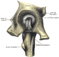
<span class="mw-page-title-main">Wing of ilium</span> Flat portion of the hip bone

The wing(ala)of ilium is the large expanded portion of the ilium, the bone which bounds the greater pelvis laterally. It presents for examination two surfaces—an external and an internal—a crest, and two borders—an anterior and a posterior.

<span class="mw-page-title-main">Intertrochanteric crest</span>

The intertrochanteric crest is a prominent bony ridge upon the posterior surface of the femur at the junction of the neck and the shaft of the femur. It extends between the greater trochanter superiorly, and the lesser trochanter inferiorly.

<span class="mw-page-title-main">Anterior inferior iliac spine</span>

The anterior inferior iliac spine (AIIS) is a bony eminence on the anterior border of the hip bone, or, more precisely, the wing of the ilium.

<span class="mw-page-title-main">Iliac crest</span> Top border of the hip

The crest of the ilium is the superior border of the wing of ilium and the superiolateral margin of the greater pelvis.

<span class="mw-page-title-main">Intertrochanteric line</span>

The intertrochanteric line is a line upon the anterior aspect of the proximal end of the femur, extending between the lesser trochanter and the greater trochanter. It is a rough, variable ridge.

<span class="mw-page-title-main">Articular capsule of the knee joint</span>

The articular capsule of the knee joint is the wide and lax joint capsule of the knee. It is thin in front and at the side, and contains the patella, ligaments, menisci, and bursae of the knee. The capsule consists of an inner synovial membrane, and an outer fibrous membrane separated by fatty deposits anteriorly and posteriorly.

<span class="mw-page-title-main">Capsule of hip joint</span>

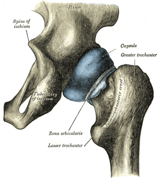
The capsule of hip joint, articular capsule, or capsular ligament is strong and dense attachment of the hip joint.

<span class="mw-page-title-main">Hip bone</span> Bone of the pelvis

The hip bone is a large flat bone, constricted in the center and expanded above and below. In some vertebrates it is composed of three parts: the ilium, ischium, and the pubis.

<span class="mw-page-title-main">Pelvis</span> Lower torso of the human body

The pelvis is the lower part of the trunk, between the abdomen and the thighs, together with its embedded skeleton.

References

PD-icon.svgThis article incorporates text in the public domain from page 335 of the 20th edition of Gray's Anatomy (1918)

  1. 1 2 Palastanga, Nigel; Soames, Roger (2012). Anatomy and Human Movement: Structure and Function. Physiotherapy Essentials (6th ed.). Edinburgh: Churchill Livingstone/Elsevier. p. 290. ISBN   978-0-7020-3553-1.
  2. Platzer, Werner (2004). Color Atlas of Human Anatomy, Vol 1: Locomotor system (5th ed.). Thieme. p. 198. ISBN   3-13-533305-1.
  3. Thieme Atlas of Anatomy. Thieme. 2006. p. 380. ISBN   3131420812.
  4. 1 2 3 4 Platzer, Werner (2004). Color Atlas of Human Anatomy, Vol 1: Locomotor system (5th ed.). Thieme. p. 200. ISBN   3-13-533305-1.