
In anatomy, the temporomandibular joints (TMJ) are the two joints connecting the jawbone to the skull. It is a bilateral synovial articulation between the temporal bone of the skull above and the condylar process of mandible below; it is from these bones that its name is derived. The joints are unique in their bilateral function, being connected via the mandible.
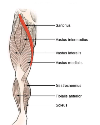
The sartorius muscle is the longest muscle in the human body. It is a long, thin, superficial muscle that runs down the length of the thigh in the anterior compartment.

The pubic symphysis is a secondary cartilaginous joint between the left and right superior rami of the pubis of the hip bones. It is in front of and below the urinary bladder. In males, the suspensory ligament of the penis attaches to the pubic symphysis. In females, the pubic symphysis is attached to the suspensory ligament of the clitoris. In most adults, it can be moved roughly 2 mm and with 1 degree rotation. This increases for women at the time of childbirth.

The piriformis muscle is a flat, pyramidally-shaped muscle in the gluteal region of the lower limbs. It is one of the six muscles in the lateral rotator group.

The acromioclavicular joint, or AC joint, is a joint at the top of the shoulder. It is the junction between the acromion and the clavicle. It is a plane synovial joint.

In vertebrate anatomy, the hip, or coxa in medical terminology, refers to either an anatomical region or a joint on the outer (lateral) side of the pelvis.

The shoulder joint is structurally classified as a synovial ball-and-socket joint and functionally as a diarthrosis and multiaxial joint. It involves an articulation between the glenoid fossa of the scapula and the head of the humerus. Due to the very loose joint capsule ,that gives a limited interface of the humerus and scapula, it is the most mobile joint of the human body.

The ischium forms the lower and back region of the hip bone.
In vertebrates, the pubis or pubic bone forms the lower and anterior part of each side of the hip bone. The pubis is the most forward-facing of the three bones that make up the hip bone. The left and right pubic bones are each made up of three sections; a superior ramus, an inferior ramus, and a body.

The fascia lata is the deep fascia of the thigh. It encloses the thigh muscles and forms the outer limit of the fascial compartments of thigh, which are internally separated by the medial intermuscular septum and the lateral intermuscular septum. The fascia lata is thickened at its lateral side where it forms the iliotibial tract, a structure that runs to the tibia and serves as a site of muscle attachment.
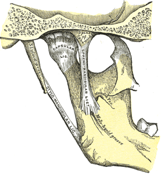
The sphenomandibular ligament is one of the three ligaments of the temporomandibular joint. It is situated medially to - and generally separate from - the articular capsule of the joint. Superiorly, it is attached to the spine of the sphenoid bone; inferiorly, it is attached to the lingula of mandible. The SML acts to limit inferior-ward movement of the mandible.

The sternoclavicular joint or sternoclavicular articulation is a synovial saddle joint between the manubrium of the sternum, and the clavicle, and the first costal cartilage. The joint possesses a joint capsule, and an articular disc, and is reinforced by multiple ligaments.

The lateral collateral ligament is an extrinsic ligament of the knee located on the lateral side of the knee. Its superior attachment is at the lateral epicondyle of the femur ; its inferior attachment is at the lateral aspect of the head of fibula. The LCL is not fused with the joint capsule. Inferiorly, the LCL splits the tendon of insertion of the biceps femoris muscle.

The iliofemoral ligament is a thick and very tough triangular capsular ligament of the hip joint situated anterior to this joint. It attaches superiorly at the inferior portion of the anterior inferior iliac spine and adjacent portion of the margin of the acetabulum; it attaches inferiorly at the intertrochanteric line.
The coracohumeral ligament is a broad ligament of the shoulder. It attaches to the coracoid process at one end, and to the greater and lesser tubercles of the humerus at the other. It strengthens the upper part of the joint capsule of the shoulder joint.
In human anatomy, the glenohumeral ligaments (GHL) are three ligaments on the anterior side of the glenohumeral joint. Reinforcing the anterior glenohumeral joint capsule, the superior, middle, and inferior glenohumeral ligaments play different roles in the stability of the head of the humerus depending on arm position and degree of rotation.
The anterior sternoclavicular ligament is a broad band of fibers attached to the clavicle above, and to the manubrium below. The ligament overlies the anterior (front) surface of sternoclavicular joint.
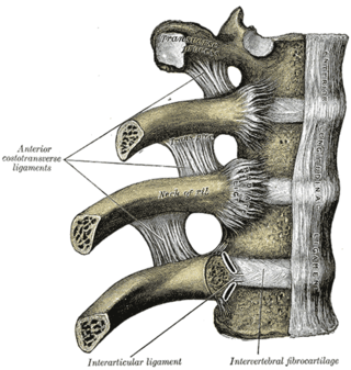
The radiate ligament of head of rib is a ligament of the costovertebral joint that typically connects the anterior edge of the head of each rib, and the side of the bodies of two adjacent vertebrae and their intervertebral discs. The ligament is formed as a thickening of the anterior portion of the joint capsule of the costovertebral joint, and thus reinforces it anteriorly.
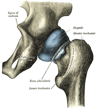
The capsule of hip joint, articular capsule, or capsular ligament is strong and dense attachment of the hip joint.

The hip bone is a large flat bone, constricted in the center and expanded above and below. In some vertebrates it is composed of three parts: the ilium, ischium, and the pubis.
