
The human leg, in the general word sense, is the entire lower limb of the human body, including the foot, thigh and even the hip or gluteal region. However, the definition in human anatomy refers only to the section of the lower limb extending from the knee to the ankle, also known as the crus. Legs are used for standing, and all forms of locomotion including recreational such as dancing, and constitute a significant portion of a person's mass. Female legs generally have greater hip anteversion and tibiofemoral angles, but shorter femur and tibial lengths than those in males.

In human anatomy, the peroneus longus is a superficial muscle in the lateral compartment of the leg, and acts to evert and plantarflex the ankle.

The tibia, also known as the shinbone or shankbone, is the larger, stronger, and anterior (frontal) of the two bones in the leg below the knee in vertebrates, and it connects the knee with the ankle bones. The tibia is found on the medial side of the leg next to the fibula and closer to the median plane or centre-line. The tibia is connected to the fibula by the interosseous membrane of the leg, forming a type of fibrous joint called a syndesmosis with very little movement. The tibia is named for the flute tibia. It is the second largest bone in the human body next to the femur. The leg bones are the strongest long bones as they support the rest of the body.

The fibula or calf bone is a leg bone on the lateral side of the tibia, to which it is connected above and below. It is the smaller of the two bones and, in proportion to its length, the slenderest of all the long bones. Its upper extremity is small, placed toward the back of the head of the tibia, below the knee joint and excluded from the formation of this joint. Its lower extremity inclines a little forward, so as to be on a plane anterior to that of the upper end; it projects below the tibia and forms the lateral part of the ankle joint.
The ankle, or the talocrural region, is the region where the foot and the leg meet. The ankle includes three joints: the ankle joint proper or talocrural joint, the subtalar joint, and the inferior tibiofibular joint. The movements produced at this joint are dorsiflexion and plantarflexion of the foot. In common usage, the term ankle refers exclusively to the ankle region. In medical terminology, "ankle" can refer broadly to the region or specifically to the talocrural joint.
Pott's fracture, also known as Pott's syndrome I and Dupuytren fracture, is an archaic term loosely applied to a variety of bimalleolar ankle fractures. The injury is caused by a combined abduction external rotation from an eversion force. This action strains the sturdy medial (deltoid) ligament of the ankle, often tearing off the medial malleolus due to its strong attachment. The talus then moves laterally, shearing off the lateral malleolus or, more commonly, breaking the fibula superior to the tibiofibular syndesmosis. If the tibia is carried anteriorly, the posterior margin of the distal end of the tibia is also sheared off by the talus. A fractured fibula in addition to detaching the medial malleolus will tear the tibiofibular syndesmosis. The combined fracture of the medial malleolus, lateral malleolus, and the posterior margin of the distal end of the tibia is known as a "trimalleolar fracture".

The tibialis anterior is a muscle in humans that originates in the upper two-thirds of the lateral (outside) surface of the tibia and inserts into the medial cuneiform and first metatarsal bones of the foot. It acts to dorsiflex and invert the foot. This muscle is mostly located near the shin.

The talus, talus bone, astragalus, or ankle bone is one of the group of foot bones known as the tarsus. The tarsus forms the lower part of the ankle joint. It transmits the entire weight of the body from the lower legs to the foot.
The Broström operation is a repair of ligaments on lateral ankle. It is designed to address ankle instability. More importantly, it is primarily used to repair the anterior talofibular ligament (ATFL) in the ankle. It is thought that the majority of patients regain most function in their ankles. The recovery time for the procedure varies according to the patient but usually takes a minimum of 3–6 months.

An ankle fracture is a break of one or more ankle bones. Symptoms may include pain, swelling, bruising, and an inability to walk on the leg. Complications may include an associated high ankle sprain, compartment syndrome, decreased range of motion, and malunion.
In human anatomy, the subtalar joint, also known as the talocalcaneal joint, is a joint of the foot. It occurs at the meeting point of the talus and the calcaneus.

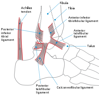
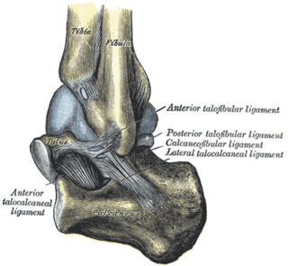
The calcaneofibular ligament is a narrow, rounded cord, running from the tip of the lateral malleolus of the fibula downward and slightly backward to a tubercle on the lateral surface of the calcaneus. It is part of the lateral collateral ligament, which opposes the hyperinversion of the subtalar joint, as in a common type of ankle sprain.
The distal tibiofibular joint is formed by the rough, convex surface of the medial side of the distal end of the fibula, and a rough concave surface on the lateral side of the tibia.
The anterior ligament of the lateral malleolus is a flat, trapezoidal band of fibers, broader below than above, which extends obliquely downward and lateralward between the adjacent margins of the tibia and fibula, on the front aspect of the syndesmosis.

The plantar calcaneonavicular ligament is a complex of three ligaments on the underside of the foot that connect the calcaneus with the navicular bone.
The deltoid ligament is a strong, flat, triangular band, attached, above, to the apex and anterior and posterior borders of the medial malleolus. The deltoid ligament is composed of: 1. Anterior tibiotalar ligament 2. Tibiocalcaneal ligament 3. Posterior tibiotalar ligament 4. Tibionavicular ligament. It consists of two sets of fibers, superficial and deep.
The posterior talofibular ligament, runs almost horizontally from the malleolar fossa of the lateral malleolus of the fibula to a prominent tubercle on the posterior surface of the talus immediately lateral to the groove for the tendon of the flexor hallucis longus.
The anterior talofibular ligament is a ligament in the ankle. It passes from the anterior margin of the fibular malleolus, anteriorly and laterally, to the talus bone, in front of its lateral articular facet. It is one of the lateral ligaments of the ankle and prevents the foot from sliding forward in relation to the shin. It is the most commonly injured ligament in a sprained ankle—from an inversion injury—and will allow a positive anterior drawer test of the ankle if completely torn.

A malleolus is the bony prominence on each side of the human ankle.
A high ankle sprain, also known as a syndesmotic ankle sprain (SAS), is a sprain of the syndesmotic ligaments that connect the tibia and fibula in the lower leg, thereby creating a mortise and tenon joint for the ankle. High ankle sprains are described as high because they are located above the ankle. They comprise approximately 15% of all ankle sprains. Unlike the common lateral ankle sprains, when ligaments around the ankle are injured through an inward twisting, high ankle sprains are caused when the lower leg and foot externally rotates.
