
In human anatomy, the wrist is variously defined as (1) the carpus or carpal bones, the complex of eight bones forming the proximal skeletal segment of the hand; (2) the wrist joint or radiocarpal joint, the joint between the radius and the carpus and; (3) the anatomical region surrounding the carpus including the distal parts of the bones of the forearm and the proximal parts of the metacarpus or five metacarpal bones and the series of joints between these bones, thus referred to as wrist joints. This region also includes the carpal tunnel, the anatomical snuff box, bracelet lines, the flexor retinaculum, and the extensor retinaculum.

The tibia, also known as the shinbone or shankbone, is the larger, stronger, and anterior (frontal) of the two bones in the leg below the knee in vertebrates ; it connects the knee with the ankle. The tibia is found on the medial side of the leg next to the fibula and closer to the median plane. The tibia is connected to the fibula by the interosseous membrane of leg, forming a type of fibrous joint called a syndesmosis with very little movement. The tibia is named for the flute tibia. It is the second largest bone in the human body, after the femur. The leg bones are the strongest long bones as they support the rest of the body.

The fibula or calf bone is a leg bone on the lateral side of the tibia, to which it is connected above and below. It is the smaller of the two bones and, in proportion to its length, the most slender of all the long bones. Its upper extremity is small, placed toward the back of the head of the tibia, below the knee joint and excluded from the formation of this joint. Its lower extremity inclines a little forward, so as to be on a plane anterior to that of the upper end; it projects below the tibia and forms the lateral part of the ankle joint.

The metatarsal bones or metatarsus are a group of five long bones in the midfoot, located between the tarsal bones and the phalanges (toes). Lacking individual names, the metatarsal bones are numbered from the medial side : the first, second, third, fourth, and fifth metatarsal. The metatarsals are analogous to the metacarpal bones of the hand. The lengths of the metatarsal bones in humans are, in descending order, second, third, fourth, fifth, and first. A bovine hind leg has two metatarsals.

The ankle, the talocrural region or the jumping bone (informal) is the area where the foot and the leg meet. The ankle includes three joints: the ankle joint proper or talocrural joint, the subtalar joint, and the inferior tibiofibular joint. The movements produced at this joint are dorsiflexion and plantarflexion of the foot. In common usage, the term ankle refers exclusively to the ankle region. In medical terminology, "ankle" can refer broadly to the region or specifically to the talocrural joint.

The lunate bone is a carpal bone in the human hand. It is distinguished by its deep concavity and crescentic outline. It is situated in the center of the proximal row carpal bones, which lie between the ulna and radius and the hand. The lunate carpal bone is situated between the lateral scaphoid bone and medial triquetral bone.

The tibialis anterior muscle is a muscle of the anterior compartment of the lower leg. It originates from the upper portion of the tibia; it inserts into the medial cuneiform and first metatarsal bones of the foot. It acts to dorsiflex and invert the foot. This muscle is mostly located near the shin.
The dorsal venous arch of the foot is a superficial vein that connects the small saphenous vein and the great saphenous vein. Anatomically, it is defined by where the dorsal veins of the first and fifth digit, respectively, meet the great saphenous vein and small saphenous vein.

The tibial nerve is a branch of the sciatic nerve. The tibial nerve passes through the popliteal fossa to pass below the arch of soleus.

The extensor digitorum longus is a pennate muscle, situated at the lateral part of the front of the leg.

The extensor digitorum brevis muscle is a muscle on the upper surface of the foot that helps extend digits 2 through 4.
In human anatomy, the fibularis brevis is a muscle that lies underneath the fibularis longus within the lateral compartment of the leg. It acts to tilt the sole of the foot away from the midline of the body (eversion) and to extend the foot downward away from the body at the ankle.

In human anatomy, the fibularis tertius is a muscle in the anterior compartment of the leg. It acts to tilt the sole of the foot away from the midline of the body (eversion) and to pull the foot upward toward the body (dorsiflexion).

The deep fibular nerve begins at the bifurcation of the common fibular nerve between the fibula and upper part of the fibularis longus, passes infero-medially, deep to the extensor digitorum longus, to the anterior surface of the interosseous membrane, and comes into relation with the anterior tibial artery above the middle of the leg; it then descends with the artery to the front of the ankle-joint, where it divides into a lateral and a medial terminal branch.

The tarsometatarsal joints are arthrodial joints in the foot. The tarsometatarsal joints involve the first, second and third cuneiform bones, the cuboid bone and the metatarsal bones. The eponym of Lisfranc joint is 18th–19th-century surgeon and gynecologist Jacques Lisfranc de St. Martin.
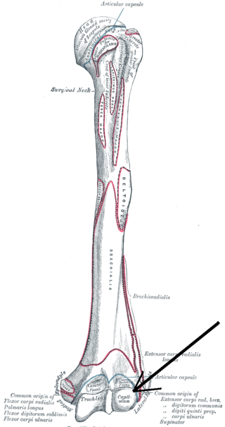
In human anatomy of the arm, the capitulum of the humerus is a smooth, rounded eminence on the lateral portion of the distal articular surface of the humerus. It articulates with the cup-shaped depression on the head of the radius, and is limited to the front and lower part of the bone.

The deltoid ligament is a strong, flat, triangular band, attached, above, to the apex and anterior and posterior borders of the medial malleolus. The deltoid ligament supports the ankle joint and also resists excessive eversion of the foot. The deltoid ligament is composed of 4 fibers:
- Anterior tibiotalar ligament
- Tibiocalcaneal ligament
- Posterior tibiotalar ligament
- Tibionavicular ligament.

The interosseous membrane of the leg extends between the interosseous crests of the tibia and fibula, helps stabilize the Tib-Fib relationship and separates the muscles on the front from those on the back of the leg.

A malleolus is the bony prominence on each side of the human ankle.

The first metatarsal bone is the bone in the foot just behind the big toe. The first metatarsal bone is the shortest of the metatarsal bones and by far the thickest and strongest of them.
 Ankle joint. Deep dissection.
 Ankle joint. Deep dissection.
