Transverse ligament of knee

Last updated
Transverse ligament of knee
Gray349.png
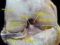
Head of right tibia seen from above, showing menisci and attachments of ligaments. (Transverse ligament labeled at upper right.)
Details
From lateral meniscus
To medial meniscus
Identifiers
Latin ligamentum transversum genus
TA98 A03.6.08.006
TA2 1889
FMA 76856
Anatomical terminology

The transverse or (anterior) meniscomeniscal ligament is a ligament in the knee joint that connects the anterior convex margin of the lateral meniscus to the anterior end of the medial meniscus.

Contents

It is divided into several strips in ten percent of subjects [1] and its thickness varies considerably in different subjects.

Function

When the knee is being extended the ligament prevents the anterior horns of the menisci from moving forward, and the condylar surfaces of the femur and tibia from exerting pressure on the menisci. [2] It has a restricting effect on anterior-posterior excursion of the anterior horn of the medial meniscus at lower degrees of knee flexion. [3]

Prevalence of meniscomeniscal ligaments

The transverse ligament is reported in 58 per cent of subjects and is thus the most prevalent of four described meniscomeniscal ligaments. The other ligaments, all three of which are reported with a frequency of less than 4 per cent, are the posterior transverse ligament, described as a bundle of fibers connecting the posterior horns of the menisci; and the medial and lateral oblique ligaments, both of which originate on the anterior horn of their namesake meniscus, passes between the cruciate ligaments, and attaches to the posterior horn of the opposite meniscus. None of the oblique ligaments have a known function. [4]

Formation

The formation of the transverse ligament has been investigated in human embryos aged 7–8 weeks (stages 18–23). At the early end of the range (stage 19), condensation of the mesenchymal interzone of the knee joint (i.e. the area of densely packed cells indicating the location of the future joint) was recognizable, and near the end of the range (stage 22) clearly visible cellular primordium of the ligament connected to both menisci was observed before all major intraarticular elements were finally evident (stage 23). [5]

Notes

  1. Platzer (2004), p 208
  2. Behnke (2006), p 197
  3. Muhle et al. (1999)
  4. Sanders et al. (1999)
  5. Ratajczak (2001), pp 323-31

Additional Images

Related Research Articles

<span class="mw-page-title-main">Human leg</span> Lower extremity or limb of the human body (foot, lower leg, thigh and hip)

The leg is the entire lower limb of the human body, including the foot, thigh or sometimes even the hip or buttock region. The major bones of the leg are the femur, tibia, and adjacent fibula. The thigh is between the hip and knee, while the calf (rear) and shin (front) are between the knee and foot.

<span class="mw-page-title-main">Knee</span> Leg joint in primates

In humans and other primates, the knee joins the thigh with the leg and consists of two joints: one between the femur and tibia, and one between the femur and patella. It is the largest joint in the human body. The knee is a modified hinge joint, which permits flexion and extension as well as slight internal and external rotation. The knee is vulnerable to injury and to the development of osteoarthritis.

<span class="mw-page-title-main">Tibia</span> Leg bone in vertebrates

The tibia, also known as the shinbone or shankbone, is the larger, stronger, and anterior (frontal) of the two bones in the leg below the knee in vertebrates ; it connects the knee with the ankle. The tibia is found on the medial side of the leg next to the fibula and closer to the median plane. The tibia is connected to the fibula by the interosseous membrane of leg, forming a type of fibrous joint called a syndesmosis with very little movement. The tibia is named for the flute tibia. It is the second largest bone in the human body, after the femur. The leg bones are the strongest long bones as they support the rest of the body.

<span class="mw-page-title-main">Segond fracture</span> Avulsion fracture of the lateral tibial condyle of the knee

The Segond fracture is a type of avulsion fracture from the lateral tibial plateau of the knee, immediately below the articular surface of the tibia.

<span class="mw-page-title-main">Meniscus (anatomy)</span> Fibrocartilaginous part of a bone joint

A meniscus is a crescent-shaped fibrocartilaginous anatomical structure that, in contrast to an articular disc, only partly divides a joint cavity. In humans, they are present in the knee, wrist, acromioclavicular, sternoclavicular, and temporomandibular joints; in other animals they may be present in other joints.

The Apley grind test or Apley test is used to evaluate individuals for problems in the meniscus of the knee. The Apley grind test has a reported sensitivity of 97% and a specificity of 87%.

A meniscus transplant or meniscal transplant is a transplant of the meniscus of the knee, which separates the thigh bone (femur) from the lower leg bone (tibia). The worn or damaged meniscus is removed and is replaced with a new one from a donor. The meniscus to be transplanted is taken from a cadaver, and, as such, is known as an allograft. Meniscal transplantation is technically difficult, as it must be sized accurately for each person, positioned properly and secured to the tibial plateau. Its success also depends on donor compatibility, stability of the transplant, and long-term health of the underlying articular cartilage.

<span class="mw-page-title-main">Medial meniscus</span> Cartilage formation in the human knee

The medial meniscus is a fibrocartilage semicircular band that spans the knee joint medially, located between the medial condyle of the femur and the medial condyle of the tibia. It is also referred to as the internal semilunar fibrocartilage. The medial meniscus has more of a crescent shape while the lateral meniscus is more circular. The anterior aspects of both menisci are connected by the transverse ligament. It is a common site of injury, especially if the knee is twisted.

<span class="mw-page-title-main">Lateral meniscus</span>

The lateral meniscus is a fibrocartilaginous band that spans the lateral side of the interior of the knee joint. It is one of two menisci of the knee, the other being the medial meniscus. It is nearly circular and covers a larger portion of the articular surface than the medial. It can occasionally be injured or torn by twisting the knee or applying direct force, as seen in contact sports.

<span class="mw-page-title-main">Trochlea of humerus</span> Articular surface of the elbow joint which articulates with the trochlear notch of the ulna

In the human arm, the humeral trochlea is the medial portion of the articular surface of the elbow joint which articulates with the trochlear notch on the ulna in the forearm.

<span class="mw-page-title-main">Lower extremity of femur</span>

The lower extremity of femur is the lower end of the femur in human and other animals, closer to the knee. It is larger than the upper extremity of femur, is somewhat cuboid in form, but its transverse diameter is greater than its antero-posterior; it consists of two oblong eminences known as the lateral condyle and medial condyle.

<span class="mw-page-title-main">Unhappy triad</span> Medical condition of the knee

The unhappy triad, also known as a blown knee among other names, is an injury to the anterior cruciate ligament, medial collateral ligament, and meniscus. Analysis during the 1990s indicated that this 'classic' O'Donoghue triad is actually an unusual clinical entity among athletes with knee injuries. Some authors mistakenly believe that in this type of injury, "combined anterior cruciate and medial collateral ligament disruptions that were incurred during athletic endeavors" always present with concomitant medial meniscus injury. However, the 1990 analysis showed that lateral meniscus tears are more common than medial meniscus tears in conjunction with sprains of the ACL.

<span class="mw-page-title-main">Articular capsule of the knee joint</span>

The articular capsule of the knee joint is the wide and lax joint capsule of the knee. It is thin in front and at the side, and contains the patella, ligaments, menisci, and bursae of the knee. The capsule consists of an inner synovial membrane, and an outer fibrous membrane separated by fatty deposits anteriorly and posteriorly.

<span class="mw-page-title-main">Coronary ligament of the knee</span>

The coronary ligaments of the knee are portions of the joint capsule which connect the inferior edges of the fibrocartilaginous menisci to the periphery of the tibial plateaus.

The anterior meniscofemoral ligament is a small fibrous band of the knee joint. It arises from the posterior horn of the lateral meniscus and passes superiorly and medially in front of the posterior cruciate ligament to attach to the lateral surface of medial condyle of the femur.

<span class="mw-page-title-main">Posterior meniscofemoral ligament</span>

The posterior meniscofemoral ligament is a small fibrous band of the knee joint. It attaches to the posterior area of the lateral meniscus and crosses superiorly and medially behind the posterior cruciate ligament to attach to the medial condyle of the femur.

<span class="mw-page-title-main">Meniscus tear</span> Rupturing of the fibrocartilage strips in the knee called menisci

A tear of a meniscus is a rupturing of one or more of the fibrocartilage strips in the knee called menisci. When doctors and patients refer to "torn cartilage" in the knee, they actually may be referring to an injury to a meniscus at the top of one of the tibiae. Menisci can be torn during innocuous activities such as walking or squatting. They can also be torn by traumatic force encountered in sports or other forms of physical exertion. The traumatic action is most often a twisting movement at the knee while the leg is bent. In older adults, the meniscus can be damaged following prolonged 'wear and tear'. Especially acute injuries can lead to displaced tears which can cause mechanical symptoms such as clicking, catching, or locking during motion of the joint. The joint will be in pain when in use, but when there is no load, the pain goes away.

<span class="mw-page-title-main">Discoid meniscus</span>

Discoid meniscus is a rare human anatomic variant that usually affects the lateral meniscus of the knee. Usually a person with this anomaly has no complaints; however, it may present as pain, swelling, or a snapping sound heard from the affected knee. Strong suggestive findings on magnetic resonance imaging includes a thickened meniscal body seen on more than two contiguous sagittal slices.

In medicine, joint locking is a symptom of pathology in a joint. It is a complaint by a person when they are unable to fully flex or fully extend a joint.

<span class="mw-page-title-main">Pelvis</span> Lower torso of the human body

The pelvis is the lower part of the trunk, between the abdomen and the thighs, together with its embedded skeleton.

References