Deltoid ligament

Last updated
Deltoid ligament
Gray354.png
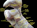
Ligaments of the medial aspect of the foot.
Details
From Talus bone (tarsal bones)
To Medial malleolus of the tibia
Identifiers
Latin ligamentum collaterale mediale articulationis talocruralis, ligamentum deltoideum
TA98 A03.6.10.003
TA2 1913
FMA 44055
Anatomical terminology

The deltoid ligament (or medial ligament of talocrural joint ) is a strong, flat, triangular band, attached, above, to the apex and anterior and posterior borders of the medial malleolus. The deltoid ligament supports the ankle joint and also resists excessive eversion of the foot. [1] The deltoid ligament is composed of 4 fibers:

Contents

  1. Anterior tibiotalar ligament
  2. Tibiocalcaneal ligament
  3. Posterior tibiotalar ligament
  4. Tibionavicular ligament.

It consists of two sets of fibers, superficial and deep.

Superficial fibres

Of the superficial fibres,

Deep fibres

The deep fibres (anterior tibiotalar) are attached from the anterior colliculus of the medial malleolus to the medial talus and medial tubercle

Coverings

The deltoid ligament is covered by the tendons of the tibialis posterior and flexor digitorum longus which are supplied by the tibial nerve (L4, L5, S1, S2, and S3).

Additional Images

Related Research Articles

<span class="mw-page-title-main">Human leg</span> Lower extremity or limb of the human body (foot, lower leg, thigh and hip)

The leg is the entire lower limb of the human body, including the foot, thigh or sometimes even the hip or buttock region. The major bones of the leg are the femur, tibia, and adjacent fibula. The thigh is between the hip and knee, while the calf (rear) and shin (front) are between the knee and foot.

<span class="mw-page-title-main">Humerus</span> Long bone of the upper arm

The humerus is a long bone in the arm that runs from the shoulder to the elbow. It connects the scapula and the two bones of the lower arm, the radius and ulna, and consists of three sections. The humeral upper extremity consists of a rounded head, a narrow neck, and two short processes. The body is cylindrical in its upper portion, and more prismatic below. The lower extremity consists of 2 epicondyles, 2 processes, and 3 fossae. As well as its true anatomical neck, the constriction below the greater and lesser tubercles of the humerus is referred to as its surgical neck due to its tendency to fracture, thus often becoming the focus of surgeons.

<span class="mw-page-title-main">Fibularis longus</span> Superficial muscle in the lateral compartment of the leg

In human anatomy, the fibularis longus is a superficial muscle in the lateral compartment of the leg. It acts to tilt the sole of the foot away from the midline of the body (eversion) and to extend the foot downward away from the body at the ankle.

<span class="mw-page-title-main">Tibia</span> Leg bone in vertebrates

The tibia, also known as the shinbone or shankbone, is the larger, stronger, and anterior (frontal) of the two bones in the leg below the knee in vertebrates ; it connects the knee with the ankle. The tibia is found on the medial side of the leg next to the fibula and closer to the median plane. The tibia is connected to the fibula by the interosseous membrane of leg, forming a type of fibrous joint called a syndesmosis with very little movement. The tibia is named for the flute tibia. It is the second largest bone in the human body, after the femur. The leg bones are the strongest long bones as they support the rest of the body.

<span class="mw-page-title-main">Fibula</span> Leg bone in vertebrates

The fibula or calf bone is a leg bone on the lateral side of the tibia, to which it is connected above and below. It is the smaller of the two bones and, in proportion to its length, the most slender of all the long bones. Its upper extremity is small, placed toward the back of the head of the tibia, below the knee joint and excluded from the formation of this joint. Its lower extremity inclines a little forward, so as to be on a plane anterior to that of the upper end; it projects below the tibia and forms the lateral part of the ankle joint.

<span class="mw-page-title-main">Ankle</span> Region where the foot and the leg meet

The ankle, the talocrural region or the jumping bone (informal) is the area where the foot and the leg meet. The ankle includes three joints: the ankle joint proper or talocrural joint, the subtalar joint, and the inferior tibiofibular joint. The movements produced at this joint are dorsiflexion and plantarflexion of the foot. In common usage, the term ankle refers exclusively to the ankle region. In medical terminology, "ankle" can refer broadly to the region or specifically to the talocrural joint.

<span class="mw-page-title-main">Calcaneus</span> Bone of the tarsus of the foot

In humans and many other primates, the calcaneus or heel bone is a bone of the tarsus of the foot which constitutes the heel. In some other animals, it is the point of the hock.

Pott's fracture, also known as Pott's syndrome I and Dupuytren fracture, is an archaic term loosely applied to a variety of bimalleolar ankle fractures. The injury is caused by a combined abduction external rotation from an eversion force. This action strains the sturdy medial (deltoid) ligament of the ankle, often tearing off the medial malleolus due to its strong attachment. The talus then moves laterally, shearing off the lateral malleolus or, more commonly, breaking the fibula superior to the tibiofibular syndesmosis. If the tibia is carried anteriorly, the posterior margin of the distal end of the tibia is also sheared off by the talus. A fractured fibula in addition to detaching the medial malleolus will tear the tibiofibular syndesmosis. The combined fracture of the medial malleolus, lateral malleolus, and the posterior margin of the distal end of the tibia is known as a "trimalleolar fracture".

<span class="mw-page-title-main">Maisonneuve fracture</span> Medical condition

The Maisonneuve fracture is a spiral fracture of the proximal third of the fibula associated with a tear of the distal tibiofibular syndesmosis and the interosseous membrane. There is an associated fracture of the medial malleolus or rupture of the deep deltoid ligament of the ankle. This type of injury can be difficult to detect.

<span class="mw-page-title-main">Talus bone</span> One of the foot bones that forms the tarsus

The talus, talus bone, astragalus, or ankle bone is one of the group of foot bones known as the tarsus. The tarsus forms the lower part of the ankle joint. It transmits the entire weight of the body from the lower legs to the foot.

<span class="mw-page-title-main">Flexor digitorum longus muscle</span> Muscle located on the tibial side of the leg

The flexor digitorum longus muscle is situated on the tibial side of the leg. At its origin it is thin and pointed, but it gradually increases in size as it descends. It serves to flex the second, third, fourth, and fifth toes.

<span class="mw-page-title-main">Subtalar joint</span>

In human anatomy, the subtalar joint, also known as the talocalcaneal joint, is a joint of the foot. It occurs at the meeting point of the talus and the calcaneus.

<span class="mw-page-title-main">Lateral meniscus</span>

The lateral meniscus is a fibrocartilaginous band that spans the lateral side of the interior of the knee joint. It is one of two menisci of the knee, the other being the medial meniscus. It is nearly circular and covers a larger portion of the articular surface than the medial. It can occasionally be injured or torn by twisting the knee or applying direct force, as seen in contact sports.

<span class="mw-page-title-main">Inferior extensor retinaculum of foot</span> Y-shaped band placed in front of the ankle-joint

The inferior extensor retinaculum of the foot is a Y-shaped band placed in front of the ankle-joint, the stem of the Y being attached laterally to the upper surface of the calcaneus, in front of the depression for the interosseous talocalcaneal ligament; it is directed medialward as a double layer, one lamina passing in front of, and the other behind, the tendons of the peroneus tertius and extensor digitorum longus.

<span class="mw-page-title-main">Anterior tibiofibular ligament</span>

The anterior ligament of the lateral malleolus is a flat, trapezoidal band of fibers, broader below than above, which extends obliquely downward and lateralward between the adjacent margins of the tibia and fibula, on the front aspect of the syndesmosis.

<span class="mw-page-title-main">Posterior longitudinal ligament</span> Ligament connecting vertebral bodies of all of the vertebrae

The posterior longitudinal ligament is a ligament connecting the posterior surfaces of the vertebral bodies of all of the vertebrae of humans. It weakly prevents hyperflexion of the vertebral column. It also prevents posterior spinal disc herniation, although problems with the ligament can cause it.

<span class="mw-page-title-main">Plantar calcaneonavicular ligament</span> Ligaments on the underside of the foot

The plantar calcaneonavicular ligament is a complex of three ligaments on the underside of the foot that connect the calcaneus with the navicular bone.

<span class="mw-page-title-main">Lateral collateral ligament of ankle joint</span> Ligaments of the ankle

The lateral collateral ligament of ankle joint are ligaments of the ankle which attach to the fibula.

<span class="mw-page-title-main">Malleolus</span> Ankle bone protrusion

A malleolus is the bony prominence on each side of the human ankle.

The posterior colliculus is the posterior portion of the medial malleolus of the distal tibia which is smaller in size comparing to the anterior colliculus. It has an attachment of the posterior tibiotalar ligament which is a part of deltoid ligament on the medial side of the ankle.

References

  1. Betts, J. Gordon (2013). Anatomy & physiology. Houston, Texas: OpenStax. ISBN   978-1-947172-04-3 . Retrieved 16 April 2023.

PD-icon.svgThis article incorporates text in the public domain from page 350 of the 20th edition of Gray's Anatomy (1918)