
The leg is the entire lower limb of the human body, including the foot, thigh or sometimes even the hip or buttock region. The major bones of the leg are the femur, tibia, and adjacent fibula.

In the human body, the cuboid bone is one of the seven tarsal bones of the foot.

In human anatomy, the dorsal interossei of the foot are four muscles situated between the metatarsal bones.
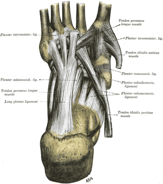
The long plantar ligament is a long ligament on the underside of the foot that connects the calcaneus with the 2nd to 5th metatarsal.
The transverse metatarsal ligament is a narrow band which runs across and connects together the heads of all the metatarsal bones. It is blended anteriorly with the plantar (glenoid) ligaments of the metatarsophalangeal articulations.

The cuneonavicular joint is a joint (articulation) in the human foot. It is formed between the navicular bone and the three cuneiform bones. The navicular and cuneiform bones are connected by dorsal and plantar ligaments.
The cuboideonavicular joint is a joint (articulation) in the foot formed between the navicular bone and cuboid bone. The navicular bone is connected with the cuboid bone by the dorsal, plantar, and interosseous cuboideonavicular ligaments. It is a syndesmosis type fibrous joint.

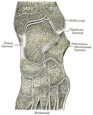
The talocalcaneonavicular joint is a ball and socket joint in the foot; the rounded head of the talus is received into the concavity formed by the posterior surface of the navicular, the anterior articular surface of the calcaneus, and the upper surface of the plantar calcaneonavicular ligament.
The calcaneocuboid joint is the joint between the calcaneus and the cuboid bone.
The plantar calcaneonavicular ligament is a complex of three ligaments on the underside of the foot that connect the calcaneus with the navicular bone.

The tarsometatarsal joints are arthrodial joints in the foot. The tarsometatarsal joints involve the first, second and third cuneiform bones, the cuboid bone and the metatarsal bones. The eponym of Lisfranc joint is 18th–19th-century surgeon and gynecologist Jacques Lisfranc de St. Martin.
The intermetatarsal joints are the articulations between the base of metatarsal bones.

The interphalangeal joints of the foot are the joints between the phalanx bones of the toes in the feet.
The intercuneiform joints are the joints the cuneiform bones.
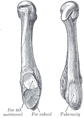
The fifth metatarsal bone is a long bone in the foot, and is palpable along the distal outer edges of the feet. It is the second smallest of the five metatarsal bones. The fifth metatarsal is analogous to the fifth metacarpal bone in the hand.

The fourth metatarsal bone is a long bone in the foot. It is smaller in size than the third metatarsal bone and is the third longest of the five metatarsal bones. The fourth metatarsal is analogous to the fourth metacarpal bone in the hand
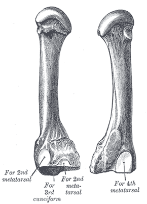
The third metatarsal bone is a long bone in the foot. It is the second longest metatarsal, the longest being the second metatarsal. The third metatarsal is analogous to the third metacarpal bone in the hand

The first metatarsal bone is the bone in the foot just behind the big toe. The first metatarsal bone is the shortest of the metatarsal bones and by far the thickest and strongest of them.
The plantar tarsometatarsal ligaments consist of longitudinal and oblique bands, disposed with less regularity than the dorsal ligaments.
In the human foot, the plantar or volar plates are fibrocartilaginous structures found in the metatarsophalangeal (MTP) and interphalangeal (IP) joints. The anatomy and composition of the plantar plates are similar to the palmar plates in the metacarpophalangeal (MCP) and interphalangeal joints in the hand; the proximal origin is thin but the distal insertion is stout. Due to the weight-bearing nature of the human foot, the plantar plates are exposed to extension forces not present in the human hand.
