The acetabulum, also called the cotyloid cavity, is a concave surface of the pelvis. The head of the femur meets with the pelvis at the acetabulum, forming the hip joint.

In vertebrate anatomy, the hip, or coxa in medical terminology, refers to either an anatomical region or a joint on the outer (lateral) side of the pelvis.

In human anatomy, the dorsal interossei of the foot are four muscles situated between the metatarsal bones.
The semimembranosus muscle is the most medial of the three hamstring muscles in the thigh. It is so named because it has a flat tendon of origin. It lies posteromedially in the thigh, deep to the semitendinosus muscle. It extends the hip joint and flexes the knee joint.

The acetabular fossa is the non-articular depressed region at the centre of the floor of the acetabulum. It is surrounded by the articular lunate surface. The floor of the fossa is formed mostly by the ischium; it is rough and thin. The space of the fossa is continuous inferiorly with the acetabular notch.

The annular ligament is a strong band of fibers that encircles the head of the radius, and retains it in contact with the radial notch of the ulna.

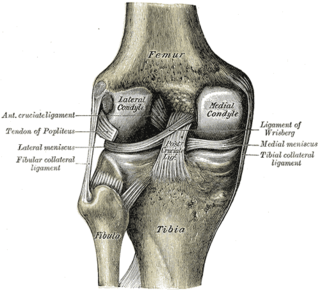
The lateral collateral ligament is an extrinsic ligament of the knee located on the lateral side of the knee. Its superior attachment is at the lateral epicondyle of the femur ; its inferior attachment is at the lateral aspect of the head of fibula. The LCL is not fused with the joint capsule. Inferiorly, the LCL splits the tendon of insertion of the biceps femoris muscle.

The iliofemoral ligament is a thick and very tough triangular capsular ligament of the hip joint situated anterior to this joint. It attaches superiorly at the inferior portion of the anterior inferior iliac spine and adjacent portion of the margin of the acetabulum; it attaches inferiorly at the intertrochanteric line.

The pubofemoral ligament is a ligament which reinforces the inferior and anterior portions of the joint capsule of the hip joint. The ligament attaches superiorly at the superior ramus of pubis, and the iliopubic eminence; it attaches inferiorly at the inferior portion of the intertrochanteric line. The psoas bursa intervenes between the ligament and joint capsule.

The ischiofemoral ligament consists of a triangular band of strong fibers on the posterior side of the hip joint. It is one of the four ligaments that reinforce the hip joint. It attaches to the posterior surface of the acetabular rim and acetabular labrum, and extends around the circumference of the joint to insert on the anterior aspect of the femur. The ischiofemoral ligament limits the internal rotation and adduction of the hip when it is in a flexed position.

The iliolumbar ligament is a strong ligament which attaches medially to the transverse process of the 5th lumbar vertebra, and laterally to back of the inner lip of the iliac crest.

The sacrococcygeal symphysis is an amphiarthrodial joint, formed between the oval surface at the apex of the sacrum, and the base of the coccyx.

The oblique popliteal ligament is a broad, flat, fibrous ligament on the posterior knee. It is an extension of the tendon of the semimembranosus muscle. It attaches onto the intercondylar fossa and lateral condyle of the femur. It reinforces the posterior central portion of the knee joint capsule.

The transverse acetabular ligament bridges the acetabular notch, creating the a foramen. The ligament is one of the sites of attachment of the ligament of head of femur.

The acetabular labrum is a fibrocartilaginous ring which surrounds the circumference of the acetabulum of the hip, deepening the acetabulum. The labrum is attached onto the bony rim and transverse acetabular ligament. It is triangular in cross-section.

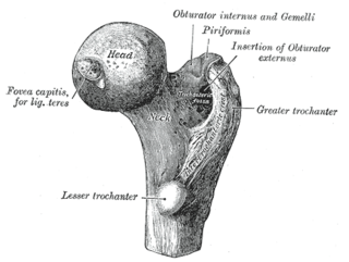
The femoral head is the highest part of the thigh bone (femur). It is supported by the femoral neck.
The arcuate popliteal ligament is an Y-shaped extracapsular ligament of the knee. It is formed as a thickening of the posterior fibres of the joint capsule of the knee. It reinforces the knee joint capsule inferolaterally.

The pelvis is the lower part of an anatomical trunk, between the abdomen and the thighs, together with its embedded skeleton.

The gemelli muscles are the inferior gemellus muscle and the superior gemellus muscle, two small accessory fasciculi to the tendon of the internal obturator muscle. The gemelli muscles belong to the lateral rotator group of six muscles of the hip that rotate the femur in the hip joint.

The lumbosacral ligament or lateral lumbosacral ligament is a ligament that helps to stabilise the lumbosacral joint. The ligament's medial attachment is at transverse process of lumbar vertebra L5; its lateral attachment is at the ala of sacrum.


