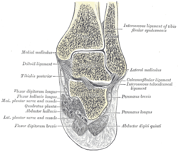
In human anatomy, the peroneus longus is a superficial muscle in the lateral compartment of the leg, and acts to evert and plantarflex the ankle.

The tibia, also known as the shinbone or shankbone, is the larger, stronger, and anterior (frontal) of the two bones in the leg below the knee in vertebrates, and it connects the knee with the ankle bones. The tibia is found on the medial side of the leg next to the fibula and closer to the median plane or centre-line. The tibia is connected to the fibula by the interosseous membrane of the leg, forming a type of fibrous joint called a syndesmosis with very little movement. The tibia is named for the flute tibia. It is the second largest bone in the human body next to the femur. The leg bones are the strongest long bones as they support the rest of the body.
The ankle, or the talocrural region, is the region where the foot and the leg meet. The ankle includes three joints: the ankle joint proper or talocrural joint, the subtalar joint, and the inferior tibiofibular joint. The movements produced at this joint are dorsiflexion and plantarflexion of the foot. In common usage, the term ankle refers exclusively to the ankle region. In medical terminology, "ankle" can refer broadly to the region or specifically to the talocrural joint.
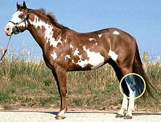
The hock, or gambrel, is the joint between the tarsal bones and tibia of a digitigrade or unguligrade quadrupedal mammal, such as a horse, cat, or dog. This joint may include articulations between tarsal bones and the fibula in some species, while in others the fibula has been greatly reduced and is only found as a vestigial remnant fused to the distal portion of the tibia. It is the anatomical homologue of the ankle of the human foot. While homologous joints occur in other tetrapods, the term is generally restricted to mammals, particularly long-legged domesticated species.
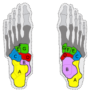
The tarsus is a cluster of seven articulating bones in each foot situated between the lower end of tibia and fibula of the lower leg and the metatarsus. It is made up of the midfoot and hindfoot.

The tibialis posterior is the most central of all the leg muscles, and is located in the deep posterior compartment of the leg.
The flexor hallucis longus muscle (FHL) is one of the three deep muscles of the posterior compartment of the leg that attaches to the plantar surface of the distal phalanx of the great toe. The other deep muscles are the flexor digitorum longus and tibialis posterior; the tibialis posterior is the most powerful of these deep muscles. All three muscles are innervated by the tibial nerve which comprises half of the sciatic nerve.

The extensor digitorum brevis muscle is a muscle on the upper surface of the foot that helps extend digits 1 through 4.
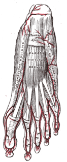
The flexor digitorum brevis lies in the middle of the sole of the foot, immediately above the central part of the plantar aponeurosis, with which it is firmly united.

The quadratus plantae is separated from the muscles of the first layer by the lateral plantar vessels and nerve. It acts to aid in flexing the 2nd to 5th toes and is one of the few muscles in the foot with no homolog in the hand.
The abductor digiti minimi is a muscle which lies along the lateral (outer) border of the foot, and is in relation by its medial margin with the lateral plantar artery, vein and nerves.

In human anatomy, the subtalar joint, also known as the talocalcaneal joint, is a joint of the foot. It occurs at the meeting point of the talus and the calcaneus.
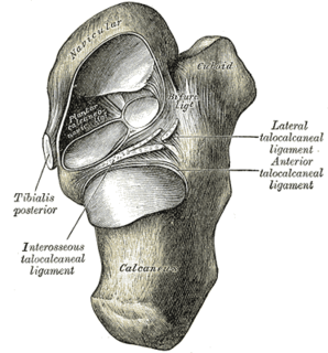
The anterior talocalcaneal ligament is a ligament in the foot.
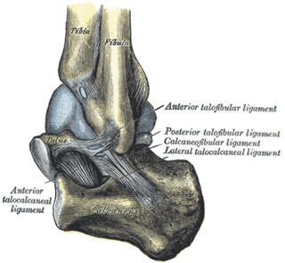
The lateral talocalcaneal ligament is a ligament in the ankle. It is a short, strong fasciculus, passing from the lateral surface of the talus, immediately beneath its fibular facet to the lateral surface of the calcaneus.
The inferior extensor retinaculum of the foot is a Y-shaped band placed in front of the ankle-joint, the stem of the Y being attached laterally to the upper surface of the calcaneus, in front of the depression for the interosseous talocalcaneal ligament; it is directed medialward as a double layer, one lamina passing in front of, and the other behind, the tendons of the peroneus tertius and extensor digitorum longus.
The talocalcaneonavicular joint is a ball and socket joint: the rounded head of the talus being received into the concavity formed by the posterior surface of the navicular, the anterior articular surface of the calcaneus, and the upper surface of the plantar calcaneonavicular ligament.

The plantar calcaneonavicular ligament is a complex of three ligaments on the underside of the foot that connect the calcaneus with the navicular bone.

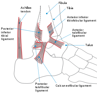
The lateral collateral ligament of ankle joint are ligaments of the ankle which attach to the fibula.
The public domain consists of all the creative works to which no exclusive intellectual property rights apply. Those rights may have expired, been forfeited, expressly waived, or may be inapplicable.

Gray's Anatomy is an English language textbook of human anatomy originally written by Henry Gray and illustrated by Henry Vandyke Carter. Earlier editions were called Anatomy: Descriptive and Surgical, Anatomy of the Human Body and Gray's Anatomy: Descriptive and Applied, but the book's name is commonly shortened to, and later editions are titled, Gray's Anatomy. The book is widely regarded as an extremely influential work on the subject, and has continued to be revised and republished from its initial publication in 1858 to the present day. The latest edition of the book, the 41st, was published in September 2015.