
The leg is the entire lower limb of the human body, including the foot, thigh or sometimes even the hip or buttock region. The major bones of the leg are the femur, tibia, and adjacent fibula. The thigh is between the hip and knee, while the calf (rear) and shin (front) are between the knee and foot.

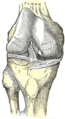
In humans and other primates, the knee joins the thigh with the leg and consists of two joints: one between the femur and tibia, and one between the femur and patella. It is the largest joint in the human body. The knee is a modified hinge joint, which permits flexion and extension as well as slight internal and external rotation. The knee is vulnerable to injury and to the development of osteoarthritis.

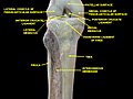
The tibia, also known as the shinbone or shankbone, is the larger, stronger, and anterior (frontal) of the two bones in the leg below the knee in vertebrates ; it connects the knee with the ankle. The tibia is found on the medial side of the leg next to the fibula and closer to the median plane. The tibia is connected to the fibula by the interosseous membrane of leg, forming a type of fibrous joint called a syndesmosis with very little movement. The tibia is named for the flute tibia. It is the second largest bone in the human body, after the femur. The leg bones are the strongest long bones as they support the rest of the body.

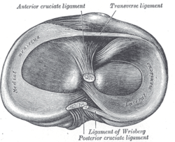
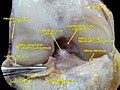
The posterior cruciate ligament (PCL) is a ligament in each knee of humans and various other animals. It works as a counterpart to the anterior cruciate ligament (ACL). It connects the posterior intercondylar area of the tibia to the medial condyle of the femur. This configuration allows the PCL to resist forces pushing the tibia posteriorly relative to the femur.
The anterior cruciate ligament (ACL) is one of a pair of cruciate ligaments in the human knee. The two ligaments are also called "cruciform" ligaments, as they are arranged in a crossed formation. In the quadruped stifle joint, based on its anatomical position, it is also referred to as the cranial cruciate ligament. The term cruciate translates to cross. This name is fitting because the ACL crosses the posterior cruciate ligament to form an "X". It is composed of strong, fibrous material and assists in controlling excessive motion. This is done by limiting mobility of the joint. The anterior cruciate ligament is one of the four main ligaments of the knee, providing 85% of the restraining force to anterior tibial displacement at 30 and 90° of knee flexion. The ACL is the most injured ligament of the four located in the knee.

The Segond fracture is a type of avulsion fracture from the lateral tibial plateau of the knee, immediately below the articular surface of the tibia.
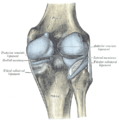
The medial collateral ligament (MCL), also called the superficial medial collateral ligament (sMCL) or tibial collateral ligament (TCL), is one of the major ligaments of the knee. It is on the medial (inner) side of the knee joint and occurs in humans and other primates. Its primary function is to resist valgus forces on the knee.

A meniscus is a crescent-shaped fibrocartilaginous anatomical structure that, in contrast to an articular disc, only partly divides a joint cavity. In humans, they are present in the knee, wrist, acromioclavicular, sternoclavicular, and temporomandibular joints; in other animals they may be present in other joints.

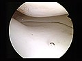
The lateral meniscus is a fibrocartilaginous band that spans the lateral side of the interior of the knee joint. It is one of two menisci of the knee, the other being the medial meniscus. It is nearly circular and covers a larger portion of the articular surface than the medial. It can occasionally be injured or torn by twisting the knee or applying direct force, as seen in contact sports.
The knee examination, in medicine and physiotherapy, is performed as part of a physical examination, or when a patient presents with knee pain or a history that suggests a pathology of the knee joint.

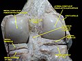
The lower extremity of femur is the lower end of the femur in human and other animals, closer to the knee. It is larger than the upper extremity of femur, is somewhat cuboid in form, but its transverse diameter is greater than its antero-posterior; it consists of two oblong eminences known as the lateral condyle and medial condyle.
The lateral collateral ligament is an extrinsic ligament of the knee located on the lateral side of the knee. Its superior attachment is at the lateral epicondyle of the femur ; its inferior attachment is at the lateral aspect of the head of fibula. The LCL is not fused with the joint capsule. Inferiorly, the LCL splits the tendon of insertion of the biceps femoris muscle.
The unhappy triad, also known as a blown knee among other names, is an injury to the anterior cruciate ligament, medial collateral ligament, and meniscus. Analysis during the 1990s indicated that this 'classic' O'Donoghue triad is actually an unusual clinical entity among athletes with knee injuries. Some authors mistakenly believe that in this type of injury, "combined anterior cruciate and medial collateral ligament disruptions that were incurred during athletic endeavors" always present with concomitant medial meniscus injury. However, the 1990 analysis showed that lateral meniscus tears are more common than medial meniscus tears in conjunction with sprains of the ACL.

The articular capsule of the knee joint is the wide and lax joint capsule of the knee. It is thin in front and at the side, and contains the patella, ligaments, menisci, and bursae of the knee. The capsule consists of an inner synovial membrane, and an outer fibrous membrane separated by fatty deposits anteriorly and posteriorly.

The Posterior meniscofemoral ligament is a small fibrous band of the knee joint. It attaches to the posterior area of the lateral meniscus and crosses superiorly and medially behind the posterior cruciate ligament to attach to the medial condyle of the femur.
A tear of a meniscus is a rupturing of one or more of the fibrocartilage strips in the knee called menisci. When doctors and patients refer to "torn cartilage" in the knee, they actually may be referring to an injury to a meniscus at the top of one of the tibiae. Menisci can be torn during innocuous activities such as walking or squatting. They can also be torn by traumatic force encountered in sports or other forms of physical exertion. The traumatic action is most often a twisting movement at the knee while the leg is bent. In older adults, the meniscus can be damaged following prolonged 'wear and tear'. Especially acute injuries can lead to displaced tears which can cause mechanical symptoms such as clicking, catching, or locking during motion of the joint. The joint will be in pain when in use, but when there is no load, the pain goes away.
The transverse or [anterior] meniscomeniscal ligament is a ligament in the knee joint that connects the anterior convex margin of the lateral meniscus to the anterior end of the medial meniscus.

Discoid meniscus is a rare human anatomic variant that usually affects the lateral meniscus of the knee. Usually a person with this anomaly has no complaints; however, it may present as pain, swelling, or a snapping sound heard from the affected knee. Strong suggestive findings on magnetic resonance imaging includes a thickened meniscal body seen on more than two contiguous sagittal slices.

A tibial plateau fracture is a break of the upper part of the tibia (shinbone) that involves the knee joint. This could involve the medial, lateral, central, or bicondylar. Symptoms include pain, swelling, and a decreased ability to move the knee. People are generally unable to walk. Complication may include injury to the artery or nerve, arthritis, and compartment syndrome.
The anterolateral ligament (ALL) is a ligament on the lateral aspect of the human knee, anterior to the fibular collateral ligament.


