
The carpal bones are the eight small bones that make up the wrist that connects the hand to the forearm. The term "carpus" is derived from the Latin carpus and the Greek καρπός (karpós), meaning "wrist". In human anatomy, the main role of the wrist is to facilitate effective positioning of the hand and powerful use of the extensors and flexors of the forearm, and the mobility of individual carpal bones increase the freedom of movements at the wrist.

In human anatomy, the wrist is variously defined as 1) the carpus or carpal bones, the complex of eight bones forming the proximal skeletal segment of the hand; (2) the wrist joint or radiocarpal joint, the joint between the radius and the carpus and; (3) the anatomical region surrounding the carpus including the distal parts of the bones of the forearm and the proximal parts of the metacarpus or five metacarpal bones and the series of joints between these bones, thus referred to as wrist joints. This region also includes the carpal tunnel, the anatomical snuff box, bracelet lines, the flexor retinaculum, and the extensor retinaculum.
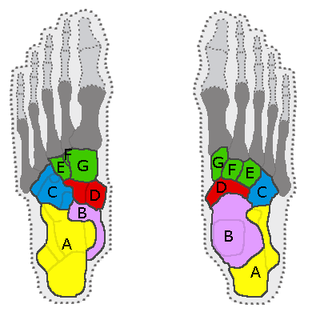
In the human body, the tarsus is a cluster of seven articulating bones in each foot situated between the lower end of the tibia and the fibula of the lower leg and the metatarsus. It is made up of the midfoot and hindfoot.

In human anatomy, the subtalar joint, also known as the talocalcaneal joint, is a joint of the foot. It occurs at the meeting point of the talus and the calcaneus.
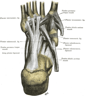
The long plantar ligament is a long ligament on the underside of the foot that connects the calcaneus with the cuboid bone.
The antebrachial fascia continuous above with the brachial fascia, is a dense, membranous investment, which forms a general sheath for the muscles in this region; it is attached, behind, to the olecranon and dorsal border of the ulna, and gives off from its deep surface numerous intermuscular septa, which enclose each muscle separately.

The sacrococcygeal symphysis is an amphiarthrodial joint, formed between the oval surface at the apex of the sacrum, and the base of the coccyx.
The posterior sacrococcygeal ligament or dorsal sacrococcygeal ligament is a ligament which stretches from the sacrum to the coccyx and thus dorsally across the sacrococcygeal symphysis shared by these two bones.

The dorsal talonavicular ligament is a broad, thin band, which connects the neck of the talus to the dorsal surface of the navicular bone; it is covered by the Extensor tendons.
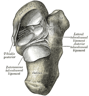
The talocalcaneonavicular joint is a ball and socket joint: the rounded head of the talus being received into the concavity formed by the posterior surface of the navicular, the anterior articular surface of the calcaneus, and the upper surface of the plantar calcaneonavicular ligament.
The intermetacarpal joints are in the hand formed between the metacarpal bones. The bases of the second, third, fourth and fifth metacarpal bones articulate with one another by small surfaces covered with cartilage. The metacarpal bones are connected together by dorsal, palmar, and interosseous ligaments.
The plantar calcaneocuboid ligament is a ligament on the bottom of the foot that connects the calcaneus to the cuboid bone. It lies deep to the long plantar ligament.

The bifurcated ligament is a strong band, attached behind to the deep hollow on the upper surface of the calcaneus and dividing in front in a Y-shaped manner into a calcaneocuboid and a calcaneonavicular part.
Intermetatarsal joints - The base of the first metatarsal is not connected with that of the second by any ligaments; in this respect the great toe resembles the thumb.

The intercuneiform joints are the joints the cuneiform bones.

The intercarpal joints can be subdivided into three sets of joints : Those of the proximal row of carpal bones, those of the distal row of carpal bones, and those of the two rows with each other.

The dorsal radioulnar ligament extends between corresponding surfaces on the dorsal aspect of the distal radioulnar articulation.
The dorsal carpometacarpal ligaments, the strongest and most distinct carpometacarpal ligaments, connect the carpal and metacarpal bones on their dorsal surfaces.
The Calcaneocuboid ligament is a fibrous band that connects the superior surface of the calcaneus to the dorsal surface of the cuboid bone.
The collateral ligaments of metatarsophalangeal joints are strong, rounded cords, placed one on either side of each joint, and attached, by one end, to the posterior tubercle on the side of the head of the metatarsal bone, and, by the other, to the contiguous extremity of the phalanx.
