Subtalar joint

Last updated
Subtalar joint
Subtalar Joint.PNG
Subtalar Joint
Gray354.png
Ligaments of the medial aspect of the foot.
Details
Identifiers
Latin articulatio subtalaris,
articulatio talocalcanea
MeSH D013380
TA98 A03.6.10.101
TA2 1924
FMA 35198
Anatomical terminology

In human anatomy, the subtalar joint, also known as the talocalcaneal joint, is a joint of the foot. It occurs at the meeting point of the talus and the calcaneus.

Contents

The joint is classed structurally as a synovial joint, [1] and functionally as a plane joint. [2]

Structure

The talus is oriented slightly obliquely on the anterior surface of the calcaneus.

There are three points of articulation between the two bones: two anteriorly and one posteriorly. The three articulations are known as facets, and they are the posterior, middle and anterior facets. [3]

The sustentaculum tali forms the floor of middle facet, and the anterior facet articulates with the head of the talus, and sits lateral and congruent to the middle facet. In some people the middle and anterior facets are joined giving just one articulation. The posterior facet is the largest of the three, and separated from the others by the tarsal canal.

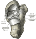
Ligaments and membranes

The main ligament of the joint is the interosseous talocalcaneal ligament, a thick, strong band of two partially joined fibers that bind the talus and calcaneus. It runs through the sinus tarsi, a canal between the articulations of the two bones.

There are four additional ligaments that form weaker connections between the talus and calcaneus.

A synovial membrane lines the capsule of the joint, and the joint is wrapped in a capsule of short fibers that are continuous with the talocalcaneonavicular and calcaneocuboid joints of the foot.

Function

The joint allows inversion and eversion of the foot, but plays minimal role in dorsiflexion or plantarflexion of the foot. [5] The centre of rotation of the subtalar joint is thought to be in the region of the middle facet. [3]

It is considered a plane synovial joint, also commonly referred to as a gliding joint. [6] It acts as a hinge connecting the talus and calcaneus. There is extensive variation in the inclination from horizontal. [7]

The subtalar joint can also be considered a combination of the anatomic subtalar joint discussed above, and also the talocalcaneal part of the talocalcaneonavicular joint. This is the more common view of the subtalar joint when discussing its movement. When both of these articulations are accounted together, it allows for pronation and supination of the midfoot to occur.

Pathology

The subtalar joint is particularly susceptible to arthritis, especially when it has previously been affected by sprains or fractures such as those of the calcaneum or talus. Symptoms of subtalar joint arthritis include pain when walking, loss of motion through the joint's range of motion, and difficulty walking on uneven surfaces. Physical therapy, orthotics, and surgery are the main treatment options.

In flat feet, the joint is typically more horizontal. [7]

Related Research Articles

<span class="mw-page-title-main">Temporomandibular joint</span> Joints connecting the jawbone to the skull

In anatomy, the temporomandibular joints (TMJ) are the two joints connecting the jawbone to the skull. It is a bilateral synovial articulation between the temporal bone of the skull above and the mandible below; it is from these bones that its name is derived. This joint is unique in that it is a bilateral joint that functions as one unit. Since the TMJ is connected to the mandible, the right and left joints must function together and therefore are not independent of each other.

<span class="mw-page-title-main">Ulna</span> Medial bone from forearm

The ulna or ulnal bone is a long bone found in the forearm that stretches from the elbow to the wrist, and when in anatomical position, is found on the medial side of the forearm. That is, the ulna is on the same side of the forearm as the little finger. It runs parallel to the radius, the other long bone in the forearm. The ulna is longer and the radius is shorter, but the radius is thicker and the ulna is thinner. Therefore, the ulna is considered to be the smaller bone of the two bones in the lower arm. The corresponding bone in the lower leg is the fibula.

<span class="mw-page-title-main">Cuboid bone</span> Bone of the ankle

In the human body, the cuboid bone is one of the seven tarsal bones of the foot.

<span class="mw-page-title-main">Trapezium (bone)</span> Bone of the wrist

The trapezium bone is a carpal bone in the hand. It forms the radial border of the carpal tunnel.

<span class="mw-page-title-main">Tibia</span> Leg bone in vertebrates

The tibia, also known as the shinbone or shankbone, is the larger, stronger, and anterior (frontal) of the two bones in the leg below the knee in vertebrates ; it connects the knee with the ankle. The tibia is found on the medial side of the leg next to the fibula and closer to the median plane. The tibia is connected to the fibula by the interosseous membrane of leg, forming a type of fibrous joint called a syndesmosis with very little movement. The tibia is named for the flute tibia. It is the second largest bone in the human body, after the femur. The leg bones are the strongest long bones as they support the rest of the body.

<span class="mw-page-title-main">Fibula</span> Leg bone in vertebrates

The fibula or calf bone is a leg bone on the lateral side of the tibia, to which it is connected above and below. It is the smaller of the two bones and, in proportion to its length, the most slender of all the long bones. Its upper extremity is small, placed toward the back of the head of the tibia, below the knee joint and excluded from the formation of this joint. Its lower extremity inclines a little forward, so as to be on a plane anterior to that of the upper end; it projects below the tibia and forms the lateral part of the ankle joint.

<span class="mw-page-title-main">Ankle</span> Region where the foot and the leg meet

The ankle, the talocrural region or the jumping bone (informal) is the area where the foot and the leg meet. The ankle includes three joints: the ankle joint proper or talocrural joint, the subtalar joint, and the inferior tibiofibular joint. The movements produced at this joint are dorsiflexion and plantarflexion of the foot. In common usage, the term ankle refers exclusively to the ankle region. In medical terminology, "ankle" can refer broadly to the region or specifically to the talocrural joint.

<span class="mw-page-title-main">Calcaneus</span> Bone of the tarsus of the foot

In humans and many other primates, the calcaneus or heel bone is a bone of the tarsus of the foot which constitutes the heel. In some other animals, it is the point of the hock.

<span class="mw-page-title-main">Radius (bone)</span> One of the two long bones of the forearm

The radius or radial bone is one of the two large bones of the forearm, the other being the ulna. It extends from the lateral side of the elbow to the thumb side of the wrist and runs parallel to the ulna. The ulna is longer than the radius, but the radius is thicker. The radius is a long bone, prism-shaped and slightly curved longitudinally.

<span class="mw-page-title-main">Talus bone</span> One of the foot bones that forms the tarsus

The talus, talus bone, astragalus, or ankle bone is one of the group of foot bones known as the tarsus. The tarsus forms the lower part of the ankle joint. It transmits the entire weight of the body from the lower legs to the foot.

<span class="mw-page-title-main">Flexor hallucis longus muscle</span> One of the three deep muscles in the lower leg

The flexor hallucis longus muscle (FHL) attaches to the plantar surface of phalanx of the great toe and is responsible for flexing that toe. The FHL is one of the three deep muscles of the posterior compartment of the leg, the others being the flexor digitorum longus and the tibialis posterior. The tibialis posterior is the most powerful of these deep muscles. All three muscles are innervated by the tibial nerve which comprises half of the sciatic nerve.

<span class="mw-page-title-main">Interosseous talocalcaneal ligament</span>

The interosseous talocalcaneal ligament forms the chief bond of union between the talus and calcaneus.

<span class="mw-page-title-main">Anterior talocalcaneal ligament</span>

The anterior talocalcaneal ligament is a ligament in the foot.

<span class="mw-page-title-main">Talocalcaneonavicular joint</span>

The talocalcaneonavicular joint is a ball and socket joint; the rounded head of the talus is received into the concavity formed by the posterior surface of the navicular, the anterior articular surface of the calcaneus, and the upper surface of the plantar calcaneonavicular ligament.

<span class="mw-page-title-main">Plantar calcaneonavicular ligament</span> Ligaments on the underside of the foot

The plantar calcaneonavicular ligament is a complex of three ligaments on the underside of the foot that connect the calcaneus with the navicular bone.

<span class="mw-page-title-main">Deltoid ligament</span> Anatomical detail in the ankle

The deltoid ligament is a strong, flat, triangular band, attached, above, to the apex and anterior and posterior borders of the medial malleolus. The deltoid ligament supports the ankle joint and also resists excessive eversion of the foot. The deltoid ligament is composed of 4 fibers:

  1. Anterior tibiotalar ligament
  2. Tibiocalcaneal ligament
  3. Posterior tibiotalar ligament
  4. Tibionavicular ligament.
<span class="mw-page-title-main">Fourth metatarsal bone</span>

The fourth metatarsal bone is a long bone in the foot. It is smaller in size than the third metatarsal bone and is the third longest of the five metatarsal bones. The fourth metatarsal is analogous to the fourth metacarpal bone in the hand

<span class="mw-page-title-main">Calcaneal fracture</span> Medical condition

A calcaneal fracture is a break of the calcaneus. Symptoms may include pain, bruising, trouble walking, and deformity of the heel. It may be associated with breaks of the hip or back.

<span class="mw-page-title-main">Outline of human anatomy</span> Overview of and topical guide to human anatomy

The following outline is provided as an overview of and topical guide to human anatomy:

<span class="mw-page-title-main">Sinus tarsi</span> Section of the hindfoot

The sinus tarsi, also known as the talocalcaneal sulcus, is a cylindrical canal in the hindfoot. It has a complex anatomy, with five ligamentous structures and a pad of adipose tissue (fat). The tarsal canal opens up into the sinus tarsi, however, the tarsal canal is a distinct structure.

References

PD-icon.svgThis article incorporates text in the public domain from page 352 of the 20th edition of Gray's Anatomy (1918)

  1. Magee, David J. (1 January 2008). Orthopedic Physical Assessment. Elsevier Health Sciences. p. 847. ISBN   978-0-7216-0571-5.
  2. Kolt, Gregory; Snyder-Mackler, Lynn (22 August 2007). Physical Therapies in Sport and Exercise. Elsevier Health Sciences. p. 421. ISBN   978-0-443-10351-3.
  3. 1 2 3 4 "Article Metrics - Centre of Rotation of the Human Subtalar Joint Using Weight-Bearing Clinical Computed Tomography | Scientific Reports". Scientific Reports. ISSN   2045-2322.
  4. http://www.physio-pedia.com/images/c/c0/Principles_of_Joint_Mobilization.pdf [ bare URL PDF ]
  5. Kyung Won, PhD. Chung (2005). Gross Anatomy (Board Review). Hagerstown, MD: Lippincott Williams & Wilkins. p. 123. ISBN   0-7817-5309-0.
  6. "The Subtalar Joint". TeachMeAnatomy. 2016-04-22. Retrieved 2018-05-07.
  7. 1 2 Michael J. Coughlin; Charles L. Saltzman; Roger A. Mann (6 September 2013). Mann's Surgery of the Foot and Ankle E-Book: Expert Consult - Online. Elsevier Health Sciences. pp. 20–22. ISBN   978-1-4557-4861-7.

Additional images