
The leg is the entire lower limb of the human body, including the foot, thigh or sometimes even the hip or buttock region. The major bones of the leg are the femur, tibia, and adjacent fibula. The thigh is between the hip and knee, while the calf (rear) and shin (front) are between the knee and foot.

In anatomy, the thigh is the area between the hip (pelvis) and the knee. Anatomically, it is part of the lower limb.
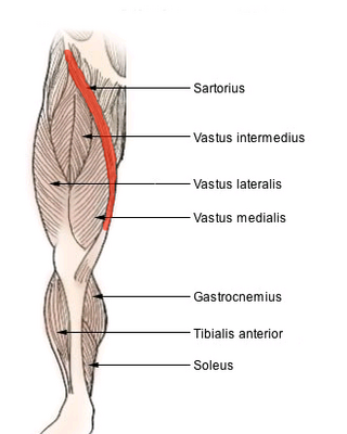
The sartorius muscle is the longest muscle in the human body. It is a long, thin, superficial muscle that runs down the length of the thigh in the anterior compartment.

The deep femoral artery also known as the deep artery of the thigh, or profunda femoris artery, is a large branch of the femoral artery. It travels more deeply ("profoundly") than the rest of the femoral artery. It gives rise to the lateral circumflex femoral artery and medial circumflex femoral artery, and the perforating arteries, terminating within the thigh.

The pectineus muscle is a flat, quadrangular muscle, situated at the anterior (front) part of the upper and medial (inner) aspect of the thigh. The pectineus muscle is the most anterior adductor of the hip. The muscle's primary action is hip flexion; it also produces adduction and internal rotation of the hip.

The external obturator muscle or obturator externus muscle is a flat, triangular muscle, which covers the outer surface of the anterior wall of the pelvis.
The adductor brevis is a muscle in the thigh situated immediately deep to the pectineus and adductor longus. It belongs to the adductor muscle group. The main function of the adductor brevis is to pull the thigh medially. The adductor brevis and the rest of the adductor muscle group is also used to stabilize left to right movements of the trunk, when standing on both feet, or to balance when standing on a moving surface. The adductor muscle group is used pressing the thighs together to ride a horse, and kicking with the inside of the foot in soccer or swimming. Last, they contribute to flexion of the thigh when running or against resistance.
In the human body, the adductor longus is a skeletal muscle located in the thigh. One of the adductor muscles of the hip, its main function is to adduct the thigh and it is innervated by the obturator nerve. It forms the medial wall of the femoral triangle.
The adductor magnus is a large triangular muscle, situated on the medial side of the thigh.
The gracilis muscle is the most superficial muscle on the medial side of the thigh. It is thin and flattened, broad above, narrow and tapering below.

The iliopsoas muscle refers to the joined psoas major and the iliacus muscles. The two muscles are separate in the abdomen, but usually merge in the thigh. They are usually given the common name iliopsoas. The iliopsoas muscle joins to the femur at the lesser trochanter. It acts as the strongest flexor of the hip.
The adductor muscles of the hip are a group of muscles in the medial compartment of the thigh mostly used for bringing the thighs together.

The lumbar plexus is a web of nerves in the lumbar region of the body which forms part of the larger lumbosacral plexus. It is formed by the divisions of the first four lumbar nerves (L1-L4) and from contributions of the subcostal nerve (T12), which is the last thoracic nerve. Additionally, the ventral rami of the fourth lumbar nerve pass communicating branches, the lumbosacral trunk, to the sacral plexus. The nerves of the lumbar plexus pass in front of the hip joint and mainly support the anterior part of the thigh.

The obturator nerve in human anatomy arises from the ventral divisions of the second, third, and fourth lumbar nerves in the lumbar plexus; the branch from the third is the largest, while that from the second is often very small.

The palatine aponeurosis a thin, firm, fibrous lamella which gives strength and support to soft palate. It serves as the insertion for the tensor veli palatini and levator veli palatini, and the origin for the musculus uvulae, palatopharyngeus, and palatoglossus.

The adductor canal is an aponeurotic tunnel in the middle third of the thigh giving passage to parts of the femoral artery, vein, and nerve. It extends from the apex of the femoral triangle to the adductor hiatus.
The posterior compartment of the thigh is one of the fascial compartments that contains the knee flexors and hip extensors known as the hamstring muscles, as well as vascular and nervous elements, particularly the sciatic nerve.

The anterior compartment of thigh contains muscles which extend the knee and flex the hip.

The deep branch of the perineal nerve is a nerve of the perineum. It is a branch of the perineal nerve, from the pudendal nerve. It supplies the superficial transverse perineal muscle, bulbospongiosus muscle, ischiocavernosus muscle, the bulb of penis, levator ani, and the external anal sphincter.
The Hannington-Kiff sign is a clinical sign in which there is an absent adductor reflex in the thigh in the presence of a positive patellar reflex. It occurs in patients with an obturator hernia, due to compression of the obturator nerve.

