Obturator nerve | |
---|---|
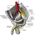
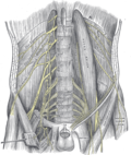
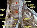
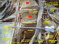
![]() Structures surrounding right hip-joint. (Obturator nerve labeled at upper right.) | |
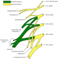
![]() Nerves of the right lower extremity. Front view. | |
Details | |
From | Lumbar plexus L2-L4 |
To | Posterior branch of obturator nerve, anterior branch of obturator nerve |
Innervates | Medial compartment of thigh |
Identifiers | |
Latin | nervus obturatorius |
MeSH | D009776 |
TA98 | A14.2.07.012 |
TA2 | 6532 |
FMA | 16487 |
Anatomical terms of neuroanatomy |
The obturator nerve in human anatomy arises from the ventral divisions of the second, third, and fourth lumbar nerves in the lumbar plexus; the branch from the third is the largest, while that from the second is often very small.
The obturator nerve originates from the anterior divisions of the L2, L3, and L4 spinal nerve roots. [1] It descends through the fibers of the psoas major, and emerges from its medial border near the brim of the pelvis. It then passes behind the common iliac arteries, and on the lateral side of the internal iliac artery and vein, and runs along the lateral wall of the lesser pelvis, above and in front of the obturator vessels, to the upper part of the obturator foramen.
Here it enters the thigh, through the obturator canal, and divides into an anterior and a posterior branch, which are separated at first by some of the fibers of the obturator externus, and lower down by the adductor brevis. [2]
An accessory obturator nerve may be present in approximately 8% to 29% of the general population. [3]
The obturator nerve is responsible for the sensory innervation of the skin of the medial aspect of the thigh.
The nerve is also responsible for the motor innervation of the adductor muscles of the lower limb (external obturator, [4] adductor longus, adductor brevis, adductor magnus, gracilis) and the pectineus (inconstant). It is, notably, not responsible for the innervation of the obturator internus, despite the similarity in name. [5]
An obturator nerve block may be used during knee surgery and urethral surgery in combination with other anaesthetics. [6]
The leg is the entire lower limb of the human body, including the foot, thigh or sometimes even the hip or buttock region. The major bones of the leg are the femur, tibia, and adjacent fibula. There are 60 bones in each leg.
A spinal nerve is a mixed nerve, which carries motor, sensory, and autonomic signals between the spinal cord and the body. In the human body there are 31 pairs of spinal nerves, one on each side of the vertebral column. These are grouped into the corresponding cervical, thoracic, lumbar, sacral and coccygeal regions of the spine. There are eight pairs of cervical nerves, twelve pairs of thoracic nerves, five pairs of lumbar nerves, five pairs of sacral nerves, and one pair of coccygeal nerves. The spinal nerves are part of the peripheral nervous system.
The pectineus muscle is a flat, quadrangular muscle, situated at the anterior (front) part of the upper and medial (inner) aspect of the thigh. The pectineus muscle is the most anterior adductor of the hip. The muscle's primary action is hip flexion; it also produces adduction and internal rotation of the hip.
The external iliac arteries are two major arteries which bifurcate off the common iliac arteries anterior to the sacroiliac joint of the pelvis.
The external obturator muscle or obturator externus muscle is a flat, triangular muscle, which covers the outer surface of the anterior wall of the pelvis.
In the human body, the adductor longus is a skeletal muscle located in the thigh. One of the adductor muscles of the hip, its main function is to adduct the thigh and it is innervated by the obturator nerve. It forms the medial wall of the femoral triangle.
The iliopsoas muscle refers to the joined psoas major and the iliacus muscles. The two muscles are separate in the abdomen, but usually merge in the thigh. They are usually given the common name iliopsoas. The iliopsoas muscle joins to the femur at the lesser trochanter. It acts as the strongest flexor of the hip.
A nerve plexus is a plexus of intersecting nerves. A nerve plexus is composed of afferent and efferent fibers that arise from the merging of the anterior rami of spinal nerves and blood vessels. There are five spinal nerve plexuses, except in the thoracic region, as well as other forms of autonomic plexuses, many of which are a part of the enteric nervous system. The nerves that arise from the plexuses have both sensory and motor functions. These functions include muscle contraction, the maintenance of body coordination and control, and the reaction to sensations such as heat, cold, pain, and pressure. There are several plexuses in the body, including:
In human anatomy, the sacral plexus is a nerve plexus which provides motor and sensory nerves for the posterior thigh, most of the lower leg and foot, and part of the pelvis. It is part of the lumbosacral plexus and emerges from the lumbar vertebrae and sacral vertebrae (L4-S4). A sacral plexopathy is a disorder affecting the nerves of the sacral plexus, usually caused by trauma, nerve compression, vascular disease, or infection. Symptoms may include pain, loss of motor control, and sensory deficits.
The internal iliac artery is the main artery of the pelvis.
The femoral nerve is a nerve in the thigh that supplies skin on the upper thigh and inner leg, and the muscles that extend the knee. It is the largest branch of the lumbar plexus.
The lumbar plexus is a web of nerves in the lumbar region of the body which forms part of the larger lumbosacral plexus. It is formed by the divisions of the first four lumbar nerves (L1-L4) and from contributions of the subcostal nerve (T12), which is the last thoracic nerve. Additionally, the ventral rami of the fourth lumbar nerve pass communicating branches, the lumbosacral trunk, to the sacral plexus. The nerves of the lumbar plexus pass in front of the hip joint and mainly support the anterior part of the thigh.
The lumbar nerves are the five pairs of spinal nerves emerging from the lumbar vertebrae. They are divided into posterior and anterior divisions.
The inferior gluteal artery is a terminal branch of the anterior trunk of the internal iliac artery. It exits the pelvis through the greater sciatic foramen. It is distributed chiefly to the buttock and the back of the thigh.
The iliac fossa is a large, smooth, concave surface on the internal surface of the ilium.
The nerve to obturator internus is a mixed nerve providing motor innervation to the obturator internus muscle and gemellus superior muscle, and sensory innervation to the hip joint. It is a branch of the sacral plexus. It is one of the group of deep gluteal nerves.
The nerve to quadratus femoris is a nerve of the sacral plexus that provides motor innervation to the quadratus femoris muscle and gemellus inferior muscle, and an articular branch to the hip joint. The nerve leaves the pelvis through the greater sciatic foramen.
The lumbosacral trunk is nervous tissue that connects the lumbar plexus with the sacral plexus. It is formed by the union of parts of the fourth and fifth lumbar nerves and descends to join the sacral plexus.
The saphenous nerve is the largest cutaneous branch of the femoral nerve. It is derived from the lumbar plexus (L3-L4). It is a strictly sensory nerve, and has no motor function. It commences in the proximal (upper) thigh and travels along the adductor canal. Upon exiting the adductor canal, the saphenous nerve terminates by splitting into two terminal branches: the sartorial nerve, and the infrapatellar nerve. The saphenous nerve is responsible for providing sensory innervation to the skin of the anteromedial leg.
The following outline is provided as an overview of and topical guide to human anatomy:
This article incorporates text in the public domain from page 953 of the 20th edition of Gray's Anatomy (1918)