Lumbar plexus

Last updated
Lumbar plexus
Lumbar plexus.svg
Plan of lumbar plexus.
Gray823.png
The lumbar plexus and its branches.
Details
From T12, L1-L4
Identifiers
Latin plexus lumbalis
plexus lumbaris
TA98 A14.2.07.002
TA2 6517
FMA 5908
Anatomical terms of neuroanatomy

The lumbar plexus is a web of nerves (a nerve plexus) in the lumbar region of the body which forms part of the larger lumbosacral plexus. It is formed by the divisions of the first four lumbar nerves (L1-L4) and from contributions of the subcostal nerve (T12), which is the last thoracic nerve. Additionally, the ventral rami of the fourth lumbar nerve pass communicating branches, the lumbosacral trunk, to the sacral plexus. The nerves of the lumbar plexus pass in front of the hip joint and mainly support the anterior part of the thigh. [1]

Contents

The plexus is formed lateral to the intervertebral foramina and passes through psoas major. Its smaller motor branches are distributed directly to psoas major, while the larger branches leave the muscle at various sites to run obliquely down through the pelvis to leave under the inguinal ligament with the exception of the obturator nerve which exits the pelvis through the obturator foramen. [1]

Branches

The iliohypogastric nerve runs posterior to the psoas major on its proximal lateral border to run laterally and obliquely on the anterior side of quadratus lumborum. Lateral to this muscle, it pierces the transversus abdominis to run above the iliac crest between that muscle and abdominal internal oblique. It gives off several motor branches to these muscles and a sensory branch to the skin of the lateral hip. Its terminal branch then runs parallel to the inguinal ligament to exit the aponeurosis of the abdominal external oblique above the external inguinal ring where it supplies the skin above the inguinal ligament (i.e. the hypogastric region) with the anterior cutaneous branch. [2]

The ilioinguinal nerve closely follows the iliohypogastric nerve on the quadratus lumborum, but then passes below it to run at the level of the iliac crest. It pierces the lateral abdominal wall and runs medially at the level of the inguinal ligament where it supplies motor branches to both transversus abdominis and sensory branches through the external inguinal ring to the skin over the pubic symphysis and the lateral aspect of the labia majora or scrotum. [2]

The genitofemoral nerve pierces psoas major anteriorly below the former two nerves to immediately split into two branches that run downward on the anterior side of the muscle. The lateral femoral branch is purely sensory. It pierces the vascular lacuna near the saphenous hiatus and supplies the skin below the inguinal ligament (i.e. proximal, lateral aspect of femoral triangle). The genital branch differs in males and females. In males it runs in the spermatic cord and in females in the inguinal canal together with the teres uteri ligament. It then sends sensory branches to the scrotal skin in males and the labia majora in females. In males it supplies motor innervation to the cremaster. [2]

The lateral cutaneous femoral nerve pierces psoas major on its lateral side and runs obliquely downward below the iliac fascia. Medial to the anterior superior iliac spine it leaves the pelvic area through the lateral muscular lacuna it enters the thigh by passing behind the lateral end of the inguinal ligament . In the thigh it briefly passes under the fascia lata before it breaches the fascia and supplies the skin of the anterior thigh. [2]

The obturator nerve leaves the lumbar plexus and descends behind psoas major on it medial side, then follows the linea terminalis into the lesser pelvis, and finally leaves the pelvic area through the obturator canal. In the thigh, it sends motor branches to obturator externus before dividing into an anterior and a posterior branch, both of which continues distally. These branches are separated by adductor brevis and supply all thigh adductors with motor innervation: pectineus, adductor longus, adductor brevis, adductor magnus, adductor minimus, and gracilis. The anterior branch contributes a terminal, sensory branch which passes along the anterior border of gracilis and supplies the skin on the medial, distal part of the thigh. [3]

The femoral nerve is the largest and longest of the plexus' nerves. It gives motor innervation to iliopsoas, pectineus, sartorius, and quadriceps femoris; and sensory innervation to the anterior thigh, posterior lower leg, and hindfoot. In the pelvic area, it runs in a groove between psoas major and iliacus giving off branches to both muscles, and exits the pelvis through the medial aspect of muscular lacuna. In the thigh it divides into numerous sensory and muscular branches and the saphenous nerve, its long sensory terminal branch which continues down to the foot. [3]

Nerves of the lumbar plexus [2]
NerveSegmentInnervated musclesCutaneous branches
Iliohypogastric T12-L1

Transversus abdominis
Abdominal internal oblique

Anterior cutaneous ramus
Lateral cutaneous ramus

Ilioinguinal L1

Anterior scrotal nerves in males
Anterior labial nerves in females

Genitofemoral L1-L2

Cremaster in males

Femoral ramus
Genital ramus

Lateral femoral cutaneous L2-L3  • Lateral femoral cutaneous
Obturator L2-L4

Obturator externus
Adductor longus
Adductor brevis
Gracilis
Pectineus
Adductor magnus

Cutaneous ramus
Femoral L2-L4

Iliacus
Pectineus
Sartorius
Quadriceps femoris

Anterior cutaneous branches
Saphenous

Direct branches from plexus to muscle
Short, direct branches L1-L3

Psoas major

 
Short, direct branches T12-L4

Quadratus lumborum
Lumbar intertransverse


Additional images

Notes

  1. 1 2 Thieme Atlas of anatomy (2006), pp 470-471
  2. 1 2 3 4 5 Thieme Atlas of anatomy (2006), pp 472-473
  3. 1 2 Thieme Atlas of anatomy (2006), pp 474-475

Related Research Articles

<span class="mw-page-title-main">Human leg</span> Lower extremity or limb of the human body (foot, lower leg, thigh and hip)

The human leg is the entire lower limb of the human body, including the foot, thigh or sometimes even the hip or buttock region. The major bones of the leg are the femur, tibia, and adjacent fibula. The thigh is between the hip and knee, while the calf (rear) and shin (front) are between the knee and foot.

<span class="mw-page-title-main">Femoral triangle</span> Anatomical region of the thigh

The femoral triangle is an anatomical region of the upper third of the thigh. It is a subfascial space which appears as a triangular depression below the inguinal ligament when the thigh is flexed, abducted and laterally rotated.

<span class="mw-page-title-main">Spinal nerve</span> Nerve that carries signals between the spinal cord and the body

A spinal nerve is a mixed nerve, which carries motor, sensory, and autonomic signals between the spinal cord and the body. In the human body there are 31 pairs of spinal nerves, one on each side of the vertebral column. These are grouped into the corresponding cervical, thoracic, lumbar, sacral and coccygeal regions of the spine. There are eight pairs of cervical nerves, twelve pairs of thoracic nerves, five pairs of lumbar nerves, five pairs of sacral nerves, and one pair of coccygeal nerves. The spinal nerves are part of the peripheral nervous system.

<span class="mw-page-title-main">Genitofemoral nerve</span>

The genitofemoral nerve is a mixed branch of the lumbar plexus derived from anterior rami of L1-L2. It splits a genital branch and a femoral branch. It provides sensory innervation to the upper anterior thigh, as well as the skin of the anterior scrotum in males and mons pubis in females. It also provides motor innervation to the cremaster muscle.

<span class="mw-page-title-main">Pectineus muscle</span> Adductor of the thigh

The pectineus muscle is a flat, quadrangular muscle, situated at the anterior (front) part of the upper and medial (inner) aspect of the thigh. The pectineus muscle is the most anterior adductor of the hip. The muscle's primary action is hip flexion; it also produces adduction and internal rotation of the hip.

<span class="mw-page-title-main">Groin</span> The two creases at the junction of the torso with the thighs, on either side of the pubic area

In human anatomy, the groin also known as the inguinal region or iliac region, is the junctional area between the torso and the thigh. The groin is at the front of the body on either side of the pubic tubercle, where the lower part of the abdominal wall meets the thigh. A fold or crease is formed at this junction known as the inguinal groove, or crease. This is also the area of the medial compartment of the thigh that contains attachments of the adductor muscles of the hip or the groin muscles. The groin is the common site for a hernia.

<span class="mw-page-title-main">Adductor longus muscle</span> Skeletal muscle located in the thigh

In the human body, the adductor longus is a skeletal muscle located in the thigh. One of the adductor muscles of the hip, its main function is to adduct the thigh and it is innervated by the obturator nerve. It forms the medial wall of the femoral triangle.

<span class="mw-page-title-main">Adductor magnus muscle</span> Muscle in the thigh

The adductor magnus is a large triangular muscle, situated on the medial side of the thigh.

<span class="mw-page-title-main">Iliopsoas</span> Joined psoas and the iliacus muscles.

The iliopsoas muscle refers to the joined psoas major and the iliacus muscles. The two muscles are separate in the abdomen, but usually merge in the thigh. They are usually given the common name iliopsoas. The iliopsoas muscle joins to the femur at the lesser trochanter. It acts as the strongest flexor of the hip.

<span class="mw-page-title-main">Nerve plexus</span> Network of nerve fibres

A nerve plexus is a plexus of intersecting nerves. A nerve plexus is composed of afferent and efferent fibers that arise from the merging of the anterior rami of spinal nerves and blood vessels. There are five spinal nerve plexuses, except in the thoracic region, as well as other forms of autonomic plexuses, many of which are a part of the enteric nervous system. The nerves that arise from the plexuses have both sensory and motor functions. These functions include muscle contraction, the maintenance of body coordination and control, and the reaction to sensations such as heat, cold, pain, and pressure. There are several plexuses in the body, including:

<span class="mw-page-title-main">Femoral nerve</span> Long nerve down the thigh and inner leg

The femoral nerve is a nerve in the thigh that supplies skin on the upper thigh and inner leg, and the muscles that extend the knee. It is the largest branch of the lumbar plexus.

<span class="mw-page-title-main">Iliohypogastric nerve</span>

The iliohypogastric nerve is a nerve that originates from the lumbar plexus that supplies sensation to skin over the lateral gluteal and hypogastric regions and motor to the internal oblique muscles and transverse abdominal muscles.

<span class="mw-page-title-main">Ilioinguinal nerve</span> Branch of the first lumbar nerve

The ilioinguinal nerve is a branch of the first lumbar nerve (L1). It separates from the first lumbar nerve along with the larger iliohypogastric nerve. It emerges from the lateral border of the psoas major just inferior to the iliohypogastric, and passes obliquely across the quadratus lumborum and iliacus. The ilioinguinal nerve then perforates the transversus abdominis near the anterior part of the iliac crest, and communicates with the iliohypogastric nerve between the transversus and the internal oblique muscle.

<span class="mw-page-title-main">Obturator nerve</span>

The obturator nerve in human anatomy arises from the ventral divisions of the second, third, and fourth lumbar nerves in the lumbar plexus; the branch from the third is the largest, while that from the second is often very small.

<span class="mw-page-title-main">Lateral cutaneous nerve of thigh</span> Nerve of the thigh

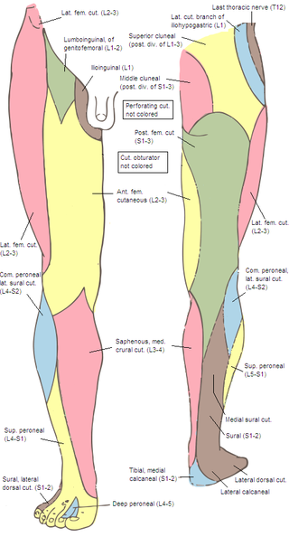
The lateral cutaneous nerve of the thigh is a cutaneous nerve of the thigh. It originates from the dorsal divisions of the second and third lumbar nerves from the lumbar plexus. It passes under the inguinal ligament to reach the thigh. It supplies sensation to the skin on the lateral part of the thigh by an anterior branch and a posterior branch.

The saphenous nerve is the largest cutaneous branch of the femoral nerve. It is derived from the lumbar plexus (L3-L4). It is a strictly sensory nerve, and has no motor function. It commences in the proximal (upper) thigh and travels along the adductor canal. Upon exiting the adductor canal, the saphenous nerve terminates by splitting into two terminal branches: the sartorial nerve, and the infrapatellar nerve. The saphenous nerve is responsible for providing sensory innervation to the skin of the anteromedial leg.

<span class="mw-page-title-main">Anterior compartment of thigh</span> Muscles which extend the knee and flex the hip

The anterior compartment of thigh contains muscles which extend the knee and flex the hip.

<span class="mw-page-title-main">Anterior cutaneous branches of the femoral nerve</span>

The anterior cutaneous branches of the femoral nerve consist of the following nerves: intermediate cutaneous nerve and medial cutaneous nerve.

<span class="mw-page-title-main">Outline of human anatomy</span> Overview of and topical guide to human anatomy

The following outline is provided as an overview of and topical guide to human anatomy:

<span class="mw-page-title-main">Pelvis</span> Lower torso of the human body

The pelvis is the lower part of the trunk, between the abdomen and the thighs, together with its embedded skeleton.

References