Scrotum

Last updated

Scrotum
Scrotum - Hodensack.JPG
Human scrotum in a relaxed state (left) and a tense state (right)
Details
Precursor Labioscrotal swelling
System Reproductive system
Artery Anterior scrotal artery, posterior scrotal artery, deep external pudendal artery, superficial external pudendal artery, internal pudendal artery
Vein Posterior scrotal veins, internal pudendal veins
Nerve Posterior scrotal nerves, anterior scrotal nerves, genital branch of genitofemoral nerve, perineal branches of posterior femoral cutaneous nerve, pudendal nerve, perineal nerve
Lymph Superficial inguinal lymph nodes
Identifiers
Latin scrotum
MeSH D012611
TA98 A09.4.03.001
A09.4.03.004
TA2 3693
FMA 18252
Anatomical terminology

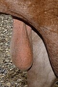
In most terrestrial mammals, the scrotum (pl.: scrotums or scrota; possibly from Latin scortum, meaning "hide" or "skin") [1] [2] or scrotal sac is a part of the external male genitalia located at the base of the penis. It consists of a sac of skin containing the external spermatic fascia, testicles, epididymides, and vasa deferentia. The scrotum will usually tighten during penile erection and when exposed to cold temperatures.

Contents

The scrotum is homologous to the labia majora in females.

Structure

Diagram of the scrotum. On the left side, the cavity of the tunica vaginalis has been opened; on the right side, only the layers superficial to the cremaster muscle have been removed. Gray1143.png
Diagram of the scrotum. On the left side, the cavity of the tunica vaginalis has been opened; on the right side, only the layers superficial to the cremaster muscle have been removed.

In regards to humans, the scrotum is a suspended dual-chambered sac of skin and muscular tissue containing the testicles and the lower part of the spermatic cords. It is located behind the penis and above the perineum. The perineal raphe is a small, vertical ridge of skin that expands from the anus and runs through the middle of the scrotum front to back. The scrotum is also a distention of the perineum and carries some abdominal tissues into its cavity including the testicular artery, testicular vein, and pampiniform plexus.

Nerve supply

NerveSurface [3]
Genital branch of genitofemoral nerve anterolateral
Anterior scrotal nerves (from ilioinguinal nerve)anterior
Posterior scrotal nerves (from perineal nerve)posterior
perineal branches of posterior femoral cutaneous nerve inferior

Blood supply

Blood vessels [4]
Anterior scrotal artery originates from the deep external pudendal artery [5]
Posterior scrotal artery
Testicular artery

Skin and glands

The skin on the scrotum is more highly pigmented in comparison to the rest of the body. The septum is a connective tissue membrane dividing the scrotum into two cavities. [6]

Lymphatic system

The scrotal lymph initially drains into the superficial inguinal lymph nodes, this then drains into the deep inguinal lymph nodes. The deep inguinal lymph nodes channel into the common iliac, which ultimately releases lymph into the cisterna chyli.

Lymphatic vessels [7]
Superficial inguinal lymph nodes
Popliteal lymph nodes

Asymmetry

One testis is typically lower than the other, which is believed to function to avoid compression in the event of impact; in humans, the left testis is typically lower than the right. [8] An alternative view is that testis descent asymmetry evolved to enable more effective cooling of the testicles. [9]

Internal structure

Image showing musculature and inner workings of the scrotum Figure 28 01 02.JPG
Image showing musculature and inner workings of the scrotum

Additional tissues and organs reside inside the scrotum and are described in more detail in the following articles:
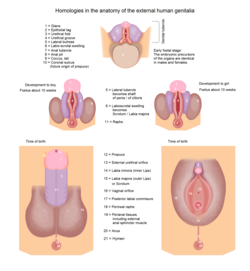
Development

Development of genitals showing homologues from indifferent to both sexes - male on left Human genitalia - development 1.png
Development of genitals showing homologues from indifferent to both sexes - male on left

During the fifth week after fertilization, the genital ridge grows behind the peritoneal membrane. By the sixth week, string-like tissues called primary sex cords form within the enlarging genital ridge. Externally, a swelling called the genital tubercule appears over the cloacal membrane.

Testosterone secretion starts during week eight, reaches peak levels during week 13 and eventually declines to very low levels by the end of the second trimester. The testosterone causes the masculinization of the labioscrotal folds into the scrotum. The scrotal raphe is formed when the embryonic, urethral groove closes by week 12. [10]

Scrotal growth and puberty

Though the testes and scrotum form early in embryonic life, sexual maturation begins upon entering puberty. The increased secretion of testosterone causes the darkening of the skin and development of pubic hair on the scrotum. [11]

Function

The scrotum regulates the temperature of the testicles and maintains it at 35 degrees Celsius (95 degrees Fahrenheit), i.e. two or three degrees below the body temperature of 37 degrees Celsius (99 degrees Fahrenheit). Higher temperatures affect spermatogenesis. [12] Temperature control is accomplished by the smooth muscles of the scrotum moving the testicles either closer to or further away from the abdomen dependent upon the ambient temperature. This is accomplished by the cremaster muscle in the abdomen and the dartos fascia (muscular tissue under the skin that makes the scrotum appear wrinkly). [11]

Having the scrotum and testicles situated outside the abdominal cavity may provide additional advantages. The external scrotum is not affected by abdominal pressure. This may prevent the emptying of the testes before the sperm were matured sufficiently for fertilization. [12] Another advantage is it protects the testes from jolts and compressions associated with an active lifestyle. The scrotum may provide some friction during intercourse, helping to enhance the activity. [13] The scrotum is also considered to be an erogenous zone. [14]

Other animals

A scrotum is present in all boreoeutherian land mammals except hippopotamuses, rhinoceroses, hedgehogs, moles, pangolins, and tapirs. The testicles remain in the body cavity in all other vertebrates, including cloacal animals. [15]

Unlike placental mammals, some male marsupials have a scrotum that is anterior to the penis, [16] [17] [18] [19] which is not homologous to the scrotum of placental mammals, [20] although there are several marsupial species without an external scrotum. [21]

The scrotum is also absent in marine mammals, such as whales, dolphins, and seals, [22] as well as in lineages of other land mammals, such as the afrotherians (elephants, aardvarks, etc.), xenarthrans (armadillos, anteaters, and sloths), and numerous families of bats and rodents. [23] [24]

Clinical significance

A study has indicated that use of a laptop computer positioned on the lap can negatively affect sperm production. [25] [26]

Diseases and conditions

The scrotum and its contents can develop many diseases and can incur injuries. These include:

See also

Bibliography

Books

Related Research Articles

<span class="mw-page-title-main">Testicle</span> Internal organ in the male reproductive system

A testicle or testis is the male gonad in all bilaterians, including humans. It is homologous to the female ovary. The functions of the testicles are to produce both sperm and androgens, primarily testosterone. Testosterone release is controlled by the anterior pituitary luteinizing hormone, whereas sperm production is controlled both by the anterior pituitary follicle-stimulating hormone and gonadal testosterone.

<span class="mw-page-title-main">Testicular torsion</span> Medical condition

Testicular torsion occurs when the spermatic cord twists, cutting off the blood supply to the testicle. The most common symptom in children is sudden, severe testicular pain. The testicle may be higher than usual in the scrotum and vomiting may occur. In newborns, pain is often absent and instead the scrotum may become discolored or the testicle may disappear from its usual place.

<span class="mw-page-title-main">Vas deferens</span> Part of the male reproductive system of many vertebrates

The vas deferens, with the more modern name ductus deferens, is part of the male reproductive system of many vertebrates. The ducts transport sperm from the epididymides to the ejaculatory ducts in anticipation of ejaculation. The vas deferens is a partially coiled tube which exits the abdominal cavity through the inguinal canal.

<span class="mw-page-title-main">Spermatic cord</span> Structure in the human male reproductive system

The spermatic cord is the cord-like structure in males formed by the vas deferens and surrounding tissue that runs from the deep inguinal ring down to each testicle. Its serosal covering, the tunica vaginalis, is an extension of the peritoneum that passes through the transversalis fascia. Each testicle develops in the lower thoracic and upper lumbar region and migrates into the scrotum. During its descent it carries along with it the vas deferens, its vessels, nerves etc. There is one on each side.

<span class="mw-page-title-main">Cryptorchidism</span> Medical condition

Cryptorchidism, also known as undescended testis, is the failure of one or both testes to descend into the scrotum. The word is from Greek κρυπτός 'hidden' and ὄρχις 'testicle'. It is the most common birth defect of the male genital tract. About 3% of full-term and 30% of premature infant boys are born with at least one undescended testis. However, about 80% of cryptorchid testes descend by the first year of life, making the true incidence of cryptorchidism around 1% overall. Cryptorchidism may develop after infancy, sometimes as late as young adulthood, but that is exceptional.

<span class="mw-page-title-main">Cremaster muscle</span> Muscle covering the testicles and spermatic cords

The cremaster muscle is a paired structure made of thin layers of striated and smooth muscle that covers the testicles and the spermatic cords in human males. It consists of the lateral and medial parts. Cremaster is an involuntary muscle, responsible for the cremasteric reflex; a protective and physiologic superficial reflex of the testicles. The reflex raises and lowers the testicles in order to keep them protected. Along with the dartos muscle of the scrotum, it regulates testicular temperature, thus aiding the process of spermatogenesis.

<span class="mw-page-title-main">Inguinal canal</span> Human abdominal anatomy

The inguinal canal is a passage in the anterior abdominal wall on each side of the body, which in males, convey the spermatic cords and in females, the round ligament of the uterus. The inguinal canals are larger and more prominent in males.

<span class="mw-page-title-main">Hydrocele testis</span> Medical condition

A hydrocele testis is an accumulation of clear fluid within the cavum vaginale, the potential space between the layers of the tunica vaginalis of the testicle. It is the most common form of hydrocele and is often referred to simply as a "hydrocele". A primary hydrocele testis causes a painless enlargement in the scrotum on the affected side and is thought to be due to the defective absorption of fluid secreted between the two layers of the tunica vaginalis. A secondary hydrocele is secondary to either inflammation or a neoplasm in the testis.

<span class="mw-page-title-main">Dartos fascia</span> Layer of tissue

The dartos fascia, dartos tunic or simply dartos is a layer of connective tissue found in the penile shaft, foreskin and scrotum. The penile portion is referred to as the superficial fascia of penis or the subcutaneous tissue of penis, while the scrotal part is the dartos proper. In addition to being continuous with itself between the scrotum and the penis, it is also continuous with Colles' fascia of the perineum and Scarpa's fascia of the abdomen.

<span class="mw-page-title-main">Hydrocele</span> Accumulation of fluid in a body cavity

A hydrocele is an accumulation of serous fluid in a body cavity. A hydrocele testis, the most common form of hydrocele, is the accumulation of fluids around a testicle. It is often caused by fluid collecting within a layer wrapped around the testicle, called the tunica vaginalis, which is derived from peritoneum. Provided there is no hernia present, it goes away without treatment in the first year. Although hydroceles usually develop in males, rare instances have been described in females in the canal of Nuck.

<span class="mw-page-title-main">Male reproductive system</span> Reproductive system of the human male

The male reproductive system consists of a number of sex organs that play a role in the process of human reproduction. These organs are located on the outside of the body, and within the pelvis.

Inguinal orchiectomy is a specific method of orchiectomy whereby one or both testicles and the full spermatic cord are surgically removed through an incision in the lower lateral abdomen. The procedure is generally performed by a urologist, typically if testicular cancer is suspected. Often it is performed as same-day surgery, with the patient returning home within hours of the procedure. Some patients elect to have a prosthetic testicle inserted into their scrotum. Depending on whether or not a prosthetic testicle is put in place of the original one, operating times run on average from three to six hours.

<span class="mw-page-title-main">Tunica vaginalis</span> Pouch of serous membrane that covers the testes

The tunica vaginalis is a pouch of serous membrane within the scrotum that lines the testis and epididymis, and the inner surface of the scrotum. It is the outermost of the three layers that constitute the capsule of the testis, with the Tunica albuginea of testis situated beneath it.

<span class="mw-page-title-main">Testicular artery</span> Branch of the abdominal aorta that supplies blood to the testicle

The testicular artery is a branch of the abdominal aorta that supplies blood to the testicle. It is a paired artery, with one for each of the testicles.

The development of the reproductive system is the part of embryonic growth that results in the sex organs and contributes to sexual differentiation. Due to its large overlap with development of the urinary system, the two systems are typically described together as the genitourinary system.

The development of the gonads is part of the prenatal development of the reproductive system and ultimately forms the testicles in males and the ovaries in females. The immature ova originate from cells from the dorsal endoderm of the yolk sac. Once they have reached the gonadal ridge they are called oogonia. Development proceeds and the oogonia become fully surrounded by a layer of connective tissue cells. In this way, the rudiments of the ovarian follicles are formed.

The reproductive system of an organism, also known as the genital system, is the biological system made up of all the anatomical organs involved in sexual reproduction. Many non-living substances such as fluids, hormones, and pheromones are also important accessories to the reproductive system. Unlike most organ systems, the sexes of differentiated species often have significant differences. These differences allow for a combination of genetic material between two individuals, which allows for the possibility of greater genetic fitness of the offspring.

<span class="mw-page-title-main">Orchiectomy</span> Surgical removal of one or both testicles

Orchiectomy is a surgical procedure in which one or both testicles are removed. The surgery can be performed for various reasons:

<span class="mw-page-title-main">Scrotal ultrasound</span> Medical ultrasound examination of the scrotum.

Scrotalultrasound is a medical ultrasound examination of the scrotum. It is used in the evaluation of testicular pain, and can help identify solid masses.

Male genital examination is a physical examination of the genital in males to detect ailments and to assess sexual development, and is normally a component of an annual physical examination. The examination includes checking the penis, scrotum, and urethral meatus. A comprehensive assessment of the male genitals assesses the pubic hair based on Sexual Maturity Rating and the size of the testicles and penis. The exam can also be conducted to verify a person's age and biological sex. The genitourinary system can also be assessed as part of the male genital examination. During a genital examination, the doctor can detect any of the following: structural abnormalities, urethral opening abnormalities, problems related to not being circumcised, lumps, tumors, redness, excoriation, edema, lesions, swelling, cancer, hair-related issues, and many others. In some instances where a physical examination of the male genitals is not sufficient to diagnose an individual, then an internal genital examination using imaging or ultrasounds will be needed for further evaluation.

References

  1. van Driel, Mels (2010). Manhood: The Rise and Fall of the Penis. Reaktion Books. p. 11. ISBN   978-1-86189-708-4 . Retrieved October 14, 2023.
  2. Spiegl, Fritz (1996). Fritz Spiegl's Sick Notes: An Alphabetical Browsing-Book of Derivatives, Abbreviations, Mnemonics and Slang for Amusement and Edification of Medics, Nurses, Patients and Hypochondriacs. Taylor & Francis. p. 142. ISBN   978-1-85070-627-4 . Retrieved October 14, 2023.
  3. Moore, Keith; Anne Agur (2007). Essential Clinical Anatomy, Third Edition. Lippincott Williams & Wilkins. p. 132. ISBN   978-0-7817-6274-8.
  4. Elson & Kapit 1977.
  5. antthigh at The Anatomy Lesson by Wesley Norman (Georgetown University)
  6. "Scrotum". Encyclopædia Britannica. Retrieved 2015-02-24.
  7. "VIII. The Lymphatic System. 5. The Lymphatics of the Lower Extremity. Gray, Henry. 1918. Anatomy of the Human Body" . Retrieved 2015-02-24.
  8. Bogaert, Anthony F. (1997). "Genital asymmetry in men" (PDF). Human Reproduction. 12 (1): 68–72. doi: 10.1093/humrep/12.1.68 . PMID   9043905. Archived from the original (PDF) on 2015-05-28. Retrieved 2015-06-29.
  9. Gallup, Gordon G.; Finn, Mary M.; Sammis, Becky (2009). "On the Origin of Descended Scrotal Testicles: The Activation Hypothesis". Evolutionary Psychology. 7 (4): 147470490900700. doi: 10.1177/147470490900700402 .
  10. Van De Graaff & Fox 1989, pp. 927–931.
  11. 1 2 Van De Graaff & Fox 1989, p. 935.
  12. 1 2 Van De Graaff & Fox 1989, p. 936.
  13. Jones, Richard (2013). Human Reproductive Biology. Academic Press. p. 74. ISBN   9780123821850. The rear-entry position of mating may allow the scrotum to stimulate the clitoris and, in this way, may produce an orgasm ...
  14. Redmon, George L. (2002). Sensual for Life. Kensington Publishing Corporation. p. 176. ISBN   978-0-75820-138-6.
  15. "Science : Bumpy lifestyle led to external testes - 17 August 1996 - New Scientist". New Scientist. Retrieved 2007-11-06.
  16. Hugh Tyndale-Biscoe; Marilyn Renfree (30 January 1987). Reproductive Physiology of Marsupials. Cambridge University Press. ISBN   978-0-521-33792-2.
  17. Libbie Henrietta Hyman (15 September 1992). Hyman's Comparative Vertebrate Anatomy. University of Chicago Press. pp. 583–. ISBN   978-0-226-87013-7.
  18. Menna Jones; Chris R. Dickman; Michael Archer (2003). Predators with Pouches: The Biology of Carnivorous Marsupials. Csiro Publishing. ISBN   978-0-643-06634-2.
  19. Norman Saunders; Lyn Hinds (1997). Marsupial Biology: Recent Research, New Perspectives. UNSW Press. ISBN   978-0-86840-311-3.
  20. Patricia J. Armati; Chris R. Dickman; Ian D. Hume (17 August 2006). Marsupials. Cambridge University Press. ISBN   978-1-139-45742-2.
  21. C. Hugh Tyndale-Biscoe (2005). Life of Marsupials. Csiro Publishing. ISBN   978-0-643-06257-3.
  22. William F. Perrin; Bernd Würsig; J.G.M. Thewissen (26 February 2009). Encyclopedia of Marine Mammals. Academic Press. ISBN   978-0-08-091993-5.
  23. "Scrotum". National Institutes of Health. Retrieved 6 January 2011.
  24. Lovegrove, B. G. (2014). "Cool sperm: Why some placental mammals have a scrotum". Journal of Evolutionary Biology. 27 (5): 801–814. doi: 10.1111/jeb.12373 . PMID   24735476. S2CID   24332311.
  25. "Laptops may damage male fertility". BBC News. 2004-12-09. Retrieved 2012-01-30.
  26. Sheynkin, Yefim; et al. (February 2005). "Increase in scrotal temperature in laptop computer users". Hum. Reprod. 20 (2): 452–455. doi: 10.1093/humrep/deh616 . PMID   15591087.
  27. "Paget's disease of the scrotum Symptoms, Diagnosis, Treatments and Causes". RightDiagnosis.com. Retrieved 2015-02-24.